| Corporate Overview August 2026 |
| 2 Forward-Looking Statements Disclaimer This presentation contains “forward-looking statements” within the meaning of the “safe harbor” provisions of the Private Securities Litigation Reform Act of 1995. Words such as “expect,” “estimate,” “project,” “budget,” “forecast,” “anticipate,” “intend,” “plan,” “may,” “will,” “could,” “should,” “believe,” “predict,” “potential,” “continue,” “strategy,” “future,” “opportunity,” “would,” “seem,” “seek,” “outlook,” “goal,” “mission,” and similar expressions are intended to identify such forward-looking statements. Forward-looking statements are predictions, projections and other statements about future events that are based on current expectations, estimates, and assumptions and, as a result, are subject to risks and uncertainties that could cause actual results to differ materially from the expected results. These statements are based on various assumptions, whether or not identified in this presentation. These forward-looking statements may include, without limitation: Zura Bio’s clinical development plans; the design, initiation, conduct, enrollment and timing of its clinical trials; expectations regarding the timing of key milestones and anticipated data readouts; expectations regarding clinical programs, including the safety, efficacy, and therapeutic potential of Zura Bio’s product candidates; expectations regarding the commercial potential of Zura Bio’s product candidates; expectations regarding data readouts from third parties, including regulatory filings and approval status; expectations regarding market opportunities, including estimated total addressable markets, competitive landscape, addressable patient populations, or potential clinical differentiation; Zura Bio’s cash resources and projected cash runway; Zura Bio’s business strategies and objectives; and any other statements that are not historical facts. These forward-looking statements are provided for illustrative purposes only and are not intended to serve as, and must not be relied on, by an investor as a guarantee, an assurance, a prediction or a definitive statement of fact or probability. Actual events are difficult or impossible to predict and could differ materially from those expressed or implied in such forward-looking statements, as a result of these risks and uncertainties, which include, but are not limited to: Zura Bio’s expectations regarding its product candidates and their related benefits, and Zura Bio’s beliefs regarding competing product candidates or approved products, may not be achieved; Zura Bio’s vision and strategy may not be successful; the timing of key events and initiation of Zura Bio’s studies, and release of clinical data may take longer than anticipated or may not be achieved at all; Zura Bio’s expectations regarding the potential general acceptability and maintenance of its product candidates by regulatory authorities, payors, physicians, and patients may not be achieved; Zura Bio’s ability to attract and retain key personnel; Zura Bio's expectations with respect to its future operating expenses, capital requirements and needs for additional financing may not be achieved; Zura Bio has not completed any clinical trials, and has no products approved for commercial sale; Zura Bio has incurred significant losses since inception, and expects to incur significant losses for the foreseeable future and may not be able to achieve or sustain profitability in the future; Zura Bio requires substantial additional capital to finance its operations, and if it is unable to raise such capital when needed or on acceptable terms, Zura Bio may be forced to delay, reduce, and/or eliminate one or more of its development programs or future commercialization efforts; Zura Bio may be unable to renew existing contracts, or enter into new contracts or may experience disputes or other challenges with respect to our vendors or other third parties; Zura Bio relies on third-party contract development manufacturing organizations for the manufacture of clinical materials; Zura Bio relies on contract research organizations, its clinical trial sites, and other third parties to conduct its preclinical studies and clinical trials; Zura Bio may be unable to obtain regulatory approval for its product candidates, and there may be related restrictions or limitations of any approved products; Zura Bio may be unable to successfully respond to general economic and geopolitical conditions; Zura Bio may be unable to effectively achieve or manage growth; Zura Bio faces competitive pressures from other companies worldwide; Zura Bio may be unable to adequately protect its intellectual property rights; and other factors set forth in documents filed, or to be filed by Zura Bio, with the Securities and Exchange Commission (SEC), including the risks and uncertainties described in the “Risk Factors” section of Zura Bio’s Annual Report on Form 10-K for the year ended December 31, 2025, and other filings with the SEC. Zura Bio cautions that the foregoing list of factors is not exclusive or exhaustive and cautions readers not to place undue reliance upon any forward-looking statements, which speak only as of the date made. Zura Bio does not undertake or accept any obligation to update any forward-looking statements, whether as a result of new information, future developments, or otherwise, except as required by law. Zura Bio gives no assurance that the expectations expressed herein will be achieved or that deviations from such expectations will not be material. This presentation discusses product candidates that are under clinical investigation and have not been approved for marketing by the U.S. Food and Drug Administration or any other regulatory authority. No representation is made as to the safety, efficacy, or likelihood of regulatory approval of these product candidates for the uses under investigation. Comparisons across clinical trials or product candidates should be interpreted with caution, as differences in study design, inclusion/exclusion criteria, patient populations, endpoints, dosing regimens, and other variables may limit interpretability. Statements in this presentation regarding clinical trials involving product candidates originating from third parties (including Eli Lilly, Pfizer and Novartis) have not been reviewed, verified, or endorsed by such parties. |
| 3 Advancing tibulizumab: a potential first- and only-in-class bispecific antibody Tibulizumab, a potential first-and only-in-class bispecific antibody inhibiting IL-17 and BAFF in development • IL-17 and BAFF among the most clinically validated pathways in immunologic disorders1,2 • Orthogonal nature of IL-17 and BAFF offers potential to modulate non-redundant pathways • Potent engagement of targets and low immunogenicity observed in Phase 1 Pipeline in a product potential: three biologically de-risked and independent shots on goal • Hidradenitis suppurativa (HS), systemic sclerosis (SSc) and polymyalgia rheumatica (PMR) span distinct biology • Yet each disease leverages existing clinical validation of the IL-17 (HS3,4 and PMR5) or BAFF arm (SSc6) with strong scientific support for the orthogonal pathway7,8,9 Zura Bio is financed beyond anticipated key near-term value inflection points • Topline data expected from HS and SSc Phase 2 studies in Q4 2026 and H1 2027, respectively • Three-asset pipeline with optionality across multiple immune-mediated indications • Cash runway through at least year end 2028 “Biologically de-risked” refers to clinical support for the relevant mechanism(s) of action and is not intended to imply regulatory or clinical success Sources: 1. Cosentyx®, Taltz®, Bimzelx® Prescribing Information.; 2. Benlysta® Prescribing Information.; 3. Kimball et al. Lancet. 2024; 4. Kimball et al. Lancet. 2023; 5. Stone et al. N Engl J Med. 2026 (REPLENISH); 6. Gordon et al. Arthritis Rheumatol. 2018; 7. Sabat et al. J Allergy Clin Immunol. 2023; 8. Lowe et al. JCI Insight. 2024; 9. van der Geest et al. Rheumatology (Oxford). 2015;10. Zura Bio filings. |
| 4 Addressing the distinct biology of immune-mediated diseases Immune-mediated diseases are often driven by multiple, intersecting immune pathways Targeting more than one pathway is a rational approach to address low or inadequate response rates in complex immune-mediated diseases Early clinical readouts across a range of immune-mediated indications have already demonstrated the potential of bispecific antibodies to break through efficacy ceilings |
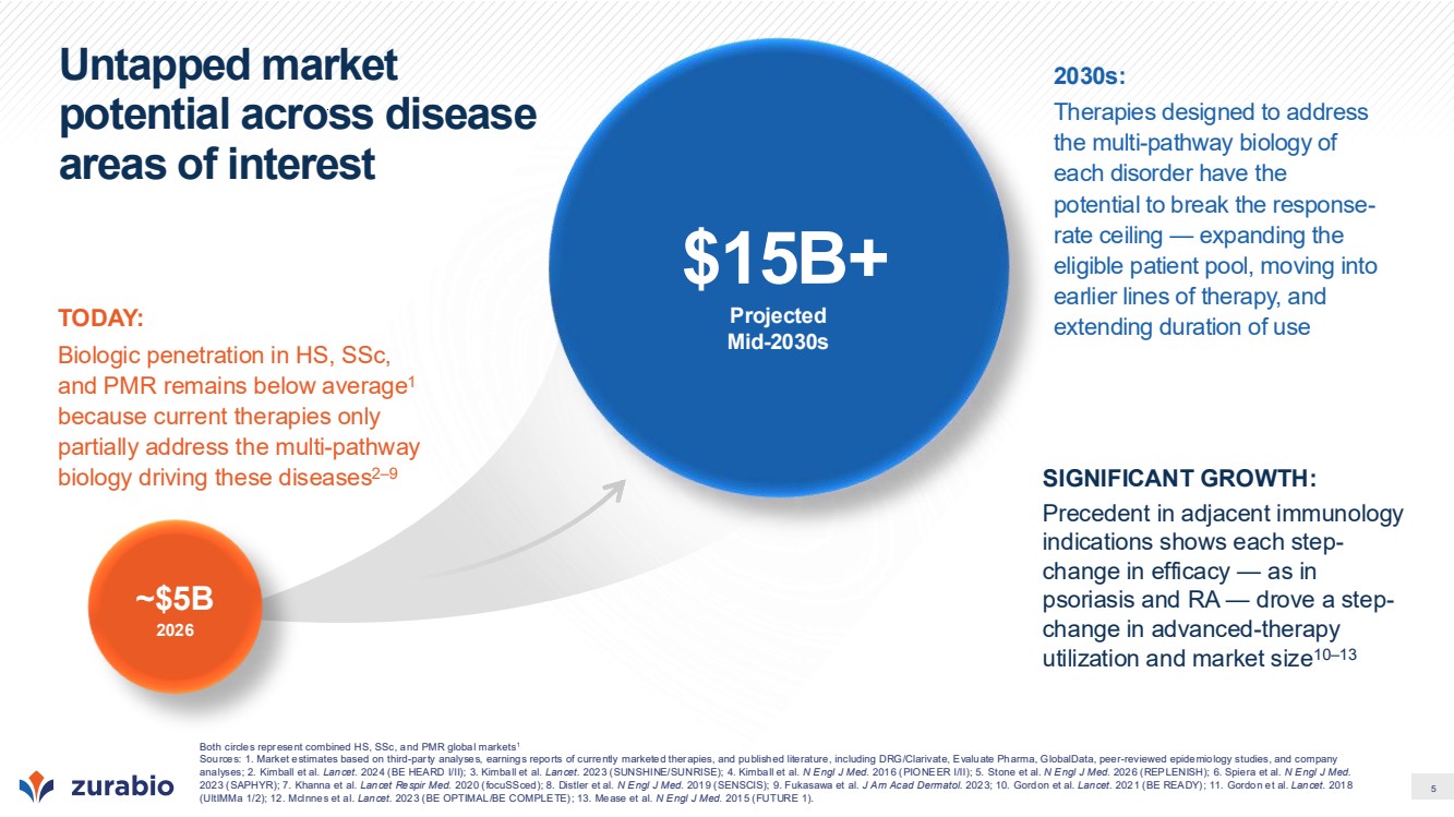
| 5 Untapped market potential across disease areas of interest ~$5B 2026 Projected Mid-2030s $15B+ TODAY: Biologic penetration in HS, SSc, and PMR remains below average1 because current therapies only partially address the multi-pathway biology driving these diseases2–9 2030s: Therapies designed to address the multi-pathway biology of each disorder have the potential to break the response-rate ceiling — expanding the eligible patient pool, moving into earlier lines of therapy, and extending duration of use SIGNIFICANT GROWTH: Precedent in adjacent immunology indications shows each step-change in efficacy — as in psoriasis and RA — drove a step-change in advanced-therapy utilization and market size10–13 Both circles represent combined HS, SSc, and PMR global markets1 Sources: 1. Market estimates based on third-party analyses, earnings reports of currently marketed therapies, and published literature, including DRG/Clarivate, Evaluate Pharma, GlobalData, peer-reviewed epidemiology studies, and company analyses; 2. Kimball et al. Lancet. 2024 (BE HEARD I/II); 3. Kimball et al. Lancet. 2023 (SUNSHINE/SUNRISE); 4. Kimball et al. N Engl J Med. 2016 (PIONEER I/II); 5. Stone et al. N Engl J Med. 2026 (REPLENISH); 6. Spiera et al. N Engl J Med. 2023 (SAPHYR); 7. Khanna et al. Lancet Respir Med. 2020 (focuSSced); 8. Distler et al. N Engl J Med. 2019 (SENSCIS); 9. Fukasawa et al. J Am Acad Dermatol. 2023; 10. Gordon et al. Lancet. 2021 (BE READY); 11. Gordon et al. Lancet. 2018 (UltIMMa 1/2); 12. McInnes et al. Lancet. 2023 (BE OPTIMAL/BE COMPLETE); 13. Mease et al. N Engl J Med. 2015 (FUTURE 1). |
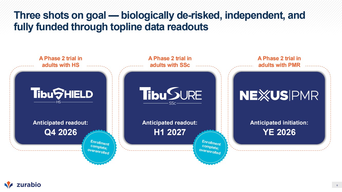
| 6 Three shots on goal — biologically de-risked, independent, and fully funded through topline data readouts A Phase 2 trial in adults with HS Anticipated readout: Q4 2026 A Phase 2 trial in adults with SSc Anticipated readout: H1 2027 A Phase 2 trial in adults with PMR Anticipated initiation: YE 2026 |
| 7 Tibulizumab: Potential first- and only-in-class therapy Tibulizumab is an investigational agent. Its efficacy and safety have not been established or approved by the FDA or any other regulatory agency worldwide. |
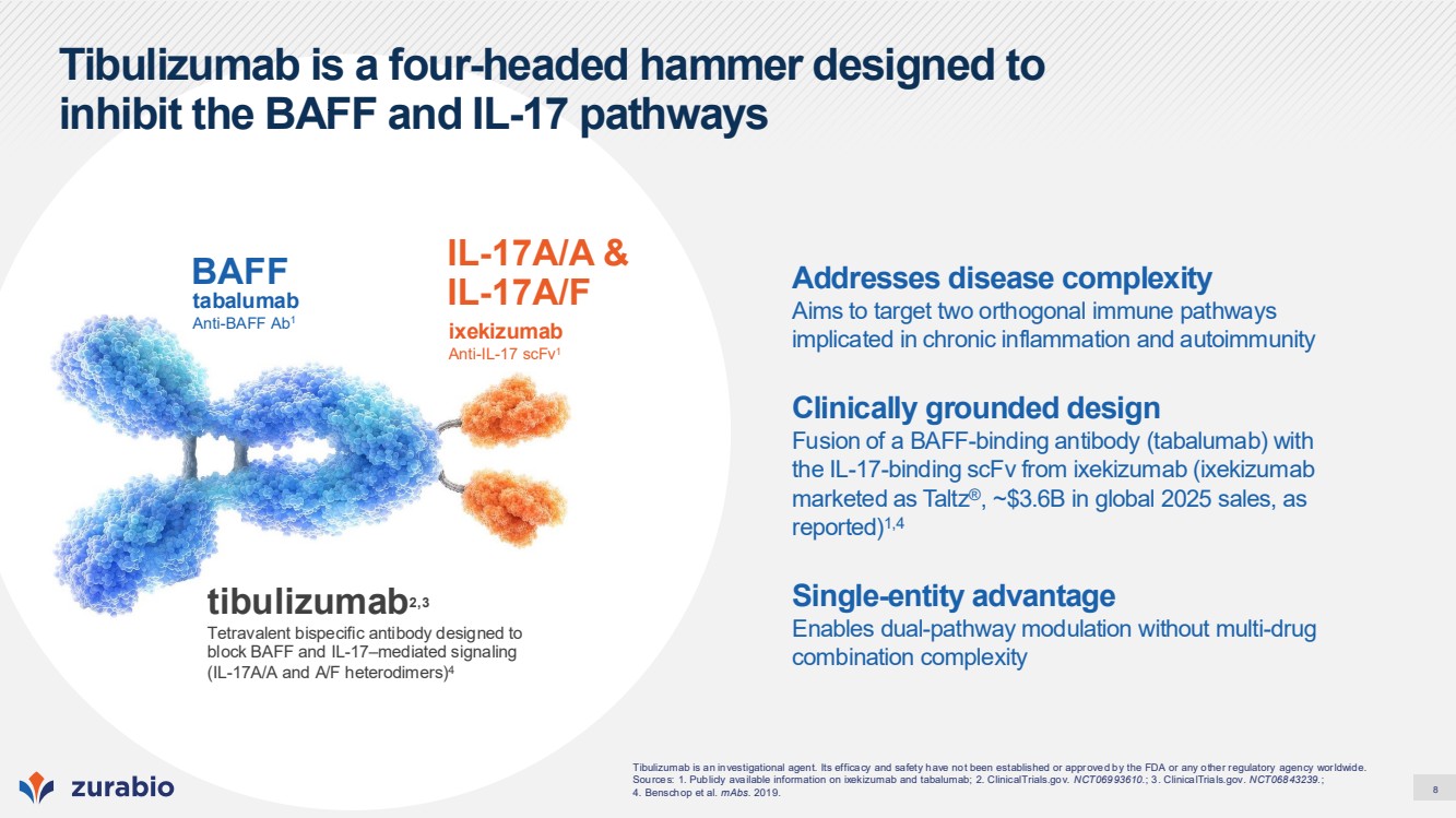
| 8 Tibulizumab is a four-headed hammer designed to inhibit the BAFF and IL-17 pathways ixekizumab Anti-IL-17 scFv1 IL-17A/A & tabalumab IL-17A/F Anti-BAFF Ab1 BAFF tibulizumab2,3 Tetravalent bispecific antibody designed to block BAFF and IL-17–mediated signaling (IL-17A/A and A/F heterodimers)4 Tibulizumab is an investigational agent. Its efficacy and safety have not been established or approved by the FDA or any other regulatory agency worldwide. Sources: 1. Publicly available information on ixekizumab and tabalumab; 2. ClinicalTrials.gov. NCT06993610.; 3. ClinicalTrials.gov. NCT06843239.; 4. Benschop et al. mAbs. 2019. Addresses disease complexity Aims to target two orthogonal immune pathways implicated in chronic inflammation and autoimmunity Clinically grounded design Fusion of a BAFF-binding antibody (tabalumab) with the IL-17-binding scFv from ixekizumab (ixekizumab marketed as Taltz®, ~$3.6B in global 2025 sales, as reported)1,4 Single-entity advantage Enables dual-pathway modulation without multi-drug combination complexity |
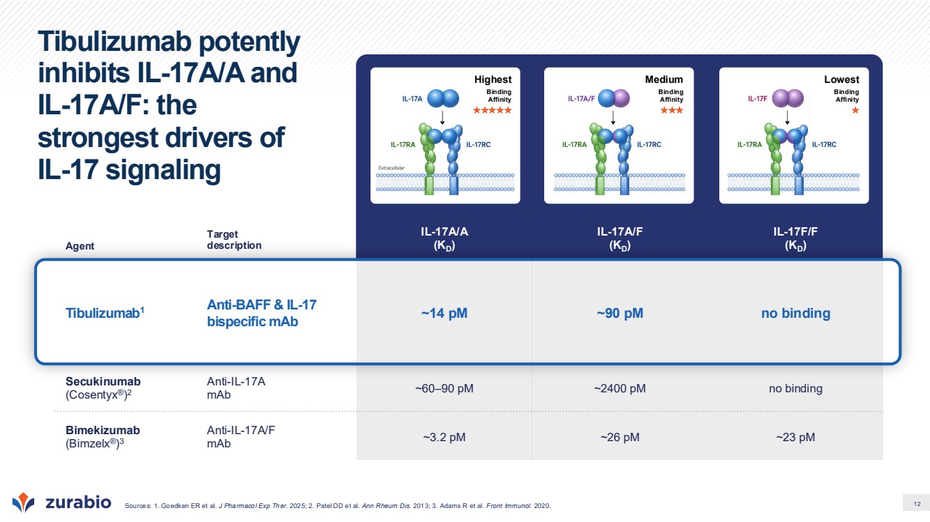
| 9 Phase 1/1b data support advancement of tibulizumab into Phase 2 development • 78 participants dosed across Phase 1/1b studies • >98% median trough target engagement for both IL-17 and BAFF in peripheral blood at 300 mg Q4W • Mean terminal half-life (t½): 27 days, supporting once-monthly dosing • Pharmacodynamic activity observed, including B cell and CRP reductions • Low incidence of treatment-emergent ADAs in multiple-dose cohorts • Safety profile consistent with published IL-17 and BAFF pathway experience, with no new or unexpected safety signals to date Time (weeks) Free BAFF concentration %CFB Free IL-17 concentration %CFB 100 mg Q4W 300 mg Q4W 100 mg Q4W 300 mg Q4W Population prediction interval* Median free target concentration Dose-dependent and comparable BAFF and IL-17 target engagement Model-based PK/PD analysis demonstrates a clear exposure–response relationship, with comparable BAFF and IL-17 suppression and near-complete target engagement at higher doses Tibulizumab is an investigational agent. Its efficacy and safety have not been established or approved by the FDA or any other regulatory agency worldwide. * Model-predicted median maximum percent change from baseline of free unbound BAFF and IL-17 following the last dose. Values shown reflect steady-state trough suppression. Eli Lilly and Company–conducted Phase 1/1b clinical studies; Zura Bio internal clinical study reports (CSRs). |
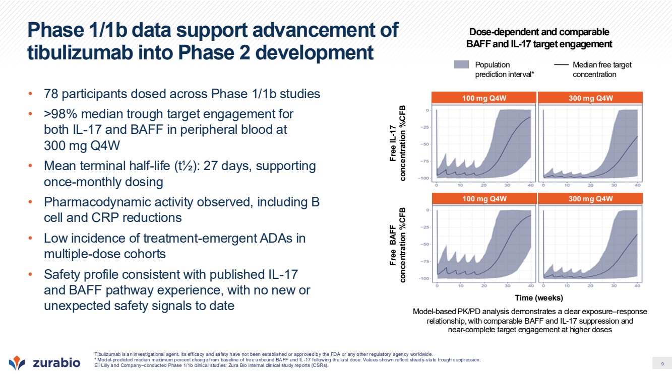
| 10 Modulation of independent innate and adaptive immune drivers of disease IL-17 Amplifies inflammation, fibrosis, and tissue destruction Drives recruitment and activation of neutrophils Enhances trafficking and cross-talk of immune cells in tissues Induces pro-inflammatory protein production, creating a self-sustaining inflammatory environment BAFF Promotes autoreactive B cell survival and function1,2,3 Promotes maturation, survival, and function of autoreactive B and plasma cells May promote chronic maintenance of lymphoid structures in tissue (TLS) Persistence of B cells leads to increased cytokines, T cell activation, and autoantibodies Broad potential in heterogeneous immune disorders Schematic representation of mechanism rationale. Not a clinical efficacy claim. References for B cell axis in HS: 1. Sabat et al. J Allergy Clin Immunol. 2022; 2. Lowe et al. JCI Insight. 2023; 3. Gudjonsson et al. JCI Insight. 2020. First and only bispecific antibody in development designed to target the orthogonal IL-17 and B cell pathways |
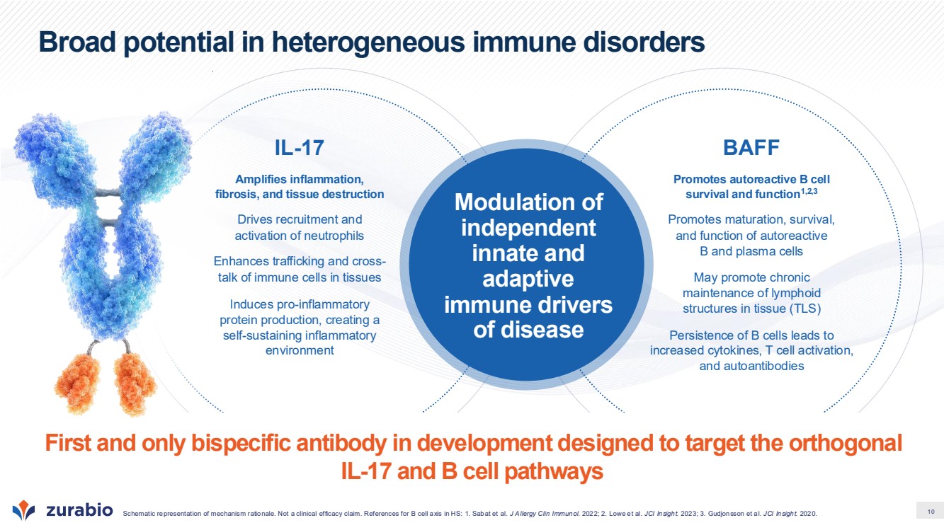
| 11 Ixekizumab contributes a high-efficacy component for IL-17 pathway inhibition • Consistent high efficacy: Demonstrated across multiple inflammatory diseases in large Phase 3 programs*1,2 • Validated IL-17 pathway inhibition: Neutralization of IL-17A/A and IL-17A/F produces robust clinical responses with a well-characterized safety profile1,2,3 • Extensive clinical safety experience: Supported by large global clinical development programs and post-marketing datasets across dermatologic and rheumatologic indications*3 • Low incidence of candidiasis relative to IL-17F/F antagonists Ixekizumab establishes a well-characterized clinical benchmark for IL-17 pathway inhibition Multiple Phase 3 programs completed High efficacy demonstrated Broad IL-17 pathway inhibition Extensive clinical and real-world experience secukinumab4 PASI 90 at Week 12* (Phase 3 psoriasis studies) PASI 90 (%; PBO-adjusted) 0 10 20 30 40 50 60 70 80 90 100 ixekizumab1,2 UNCOVER-1 UNCOVER-2 UNCOVER-3 PsO1 PsO2 * PASI 90 values represent placebo-adjusted response rates at Week 12 from independent Phase 3 psoriasis trials. Studies were not designed for head-to-head comparison; trial designs, patient populations, dosing regimens, and placebo response rates differed. Clinical efficacy data shown are limited to psoriasis studies. Efficacy and durability may vary by indication Sources: 1. Griffiths CE et al. Lancet. 2015; 2. Gordon KB et al. N Engl J Med. 2016; 3. Eli Lilly and Company and Novartis. Public disclosures and prescribing information.; 4. Langley RG et al. N Engl J Med. 2014 |
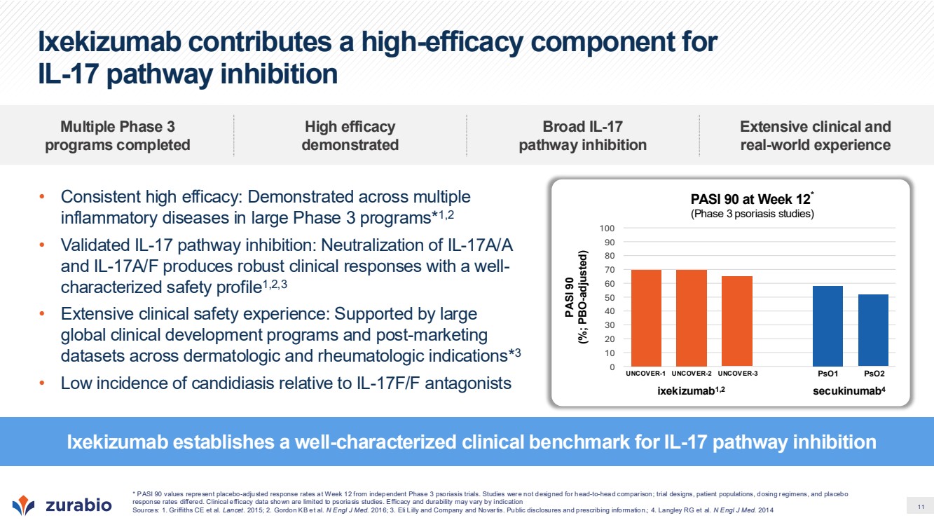
| 12 Tibulizumab potently inhibits IL-17A/A and IL-17A/F: the strongest drivers of IL-17 signaling Agent Target description IL-17A/A (KD) IL-17A/F (KD) IL-17F/F (KD) Tibulizumab1 Anti-BAFF & IL-17 bispecific mAb ~14 pM ~90 pM no binding Secukinumab (Cosentyx®) 2 Anti-IL-17A mAb ~60–90 pM ~2400 pM no binding Bimekizumab (Bimzelx®) 3 Anti-IL-17A/F mAb ~3.2 pM ~26 pM ~23 pM Sources: 1. Goedken ER et al. J Pharmacol Exp Ther. 2025; 2. Patel DD et al. Ann Rheum Dis. 2013; 3. Adams R et al. Front Immunol. 2020. Highest Binding Affinity Medium Binding Affinity Lowest Binding Affinity |
| 13 Hidradenitis Suppurativa (HS) |
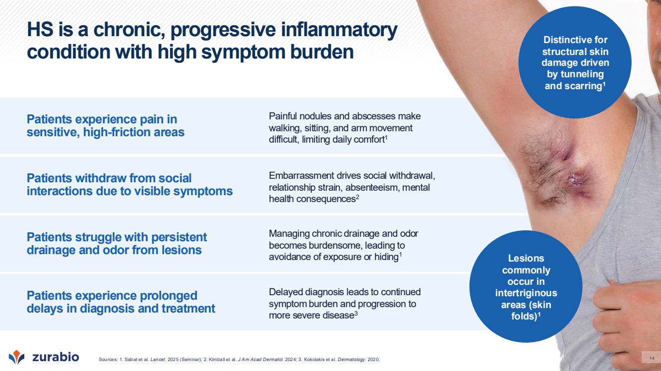
| 14 HS is a chronic, progressive inflammatory condition with high symptom burden Patients experience pain in sensitive, high-friction areas Patients withdraw from social interactions due to visible symptoms Patients struggle with persistent drainage and odor from lesions Patients experience prolonged delays in diagnosis and treatment Painful nodules and abscesses make walking, sitting, and arm movement difficult, limiting daily comfort1 Managing chronic drainage and odor becomes burdensome, leading to avoidance of exposure or hiding1 Embarrassment drives social withdrawal, relationship strain, absenteeism, mental health consequences2 Delayed diagnosis leads to continued symptom burden and progression to more severe disease3 Lesions commonly occur in intertriginous areas (skin folds)1 Distinctive for structural skin damage driven by tunneling and scarring1 Sources: 1. Sabat et al. 14 Lancet. 2025 (Seminar); 2. Kimball et al. J Am Acad Dermatol. 2024; 3. Kokolakis et al. Dermatology. 2020. |
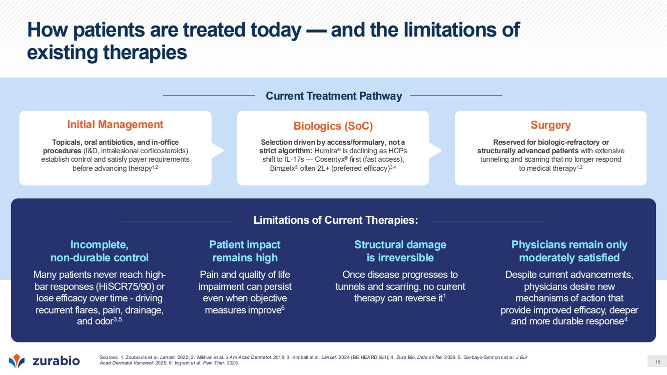
| 15 How patients are treated today — and the limitations of existing therapies Current Treatment Pathway Initial Management Topicals, oral antibiotics, and in-office procedures (I&D, intralesional corticosteroids) establish control and satisfy payer requirements before advancing therapy1,2 Biologics (SoC) Selection driven by access/formulary, not a strict algorithm: Humira® is declining as HCPs shift to IL-17s — Cosentyx® first (fast access), Bimzelx® often 2L+ (preferred efficacy)3,4 Surgery Reserved for biologic-refractory or structurally advanced patients with extensive tunneling and scarring that no longer respond to medical therapy1,2 Limitations of Current Therapies: Incomplete, non-durable control Many patients never reach high-bar responses (HiSCR75/90) or lose efficacy over time - driving recurrent flares, pain, drainage, and odor3,5 Structural damage is irreversible Once disease progresses to tunnels and scarring, no current therapy can reverse it1 Patient impact remains high Pain and quality of life impairment can persist even when objective measures improve6 Physicians remain only moderately satisfied Despite current advancements, physicians desire new mechanisms of action that provide improved efficacy, deeper and more durable response4 Sources: 1. Zouboulis et al. Lancet. 2025; 2. Alikhan et al. J Am Acad Dermatol. 2019; 3. Kimball et al. Lancet. 2024 (BE HEARD I&II); 4. Zura Bio. Data on file. 2026; 5. Garbayo-Salmons et al. J Eur Acad Dermatol Venereol. 2025; 6. Ingram et al. Pain Ther. 2025. |
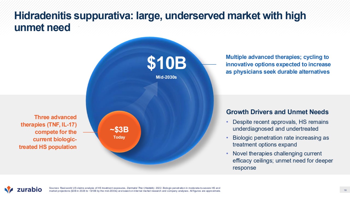
| 16 Hidradenitis suppurativa: large, underserved market with high unmet need Three advanced therapies (TNF, IL-17) compete for the current biologic-treated HS population Multiple advanced therapies; cycling to innovative options expected to increase as physicians seek durable alternatives Sources: Real-world US claims analysis of HS treatment exposures, Dermatol Ther (Heidelb). 2022. Biologic penetration in moderate-to-severe HS and market projections ($3B in 2026 to ~$10B by the mid-2030s) are based on internal market research and company analyses. All figures are approximate. Mid-2030s $10B Growth Drivers and Unmet Needs • Despite recent approvals, HS remains underdiagnosed and undertreated • Biologic penetration rate increasing as treatment options expand • Novel therapies challenging current efficacy ceilings; unmet need for deeper response ~$3B Today |
| 17 Unlike healthy skin, B cells infiltrate and chronically persist in HS lesions, driving a local amplified and targeted immune response Sources: 1. Gudjonsson et al. JCI Insight. 2020; 2. Lowe et al. JCI Insight. 2020; 3. Lowe et al. JCI Insight. 2024; 4. Yu et al. Immunity. 2024. In severe disease, B and T cells organize and persist in tertiary lymphoid structures (TLS)3,4 B cells T cells Proliferating Cells Immunofluorescence analysis of HS chronic lesion with tunnel B cells expand with disease progression2 Flow cytometry analysis of healthy skin biopsies, active HS biopsies (inflammatory nodules), or end stage HS tissue (tunnel from surgical excision) B cells and plasma cells make up the bulk of HS cellular infiltrate1 Imaging CyTOF analysis of HS lesions Plasma cells B cells Monocytes/ macrophages CD8 T cells Neutrophils Number of cells per mm2 |
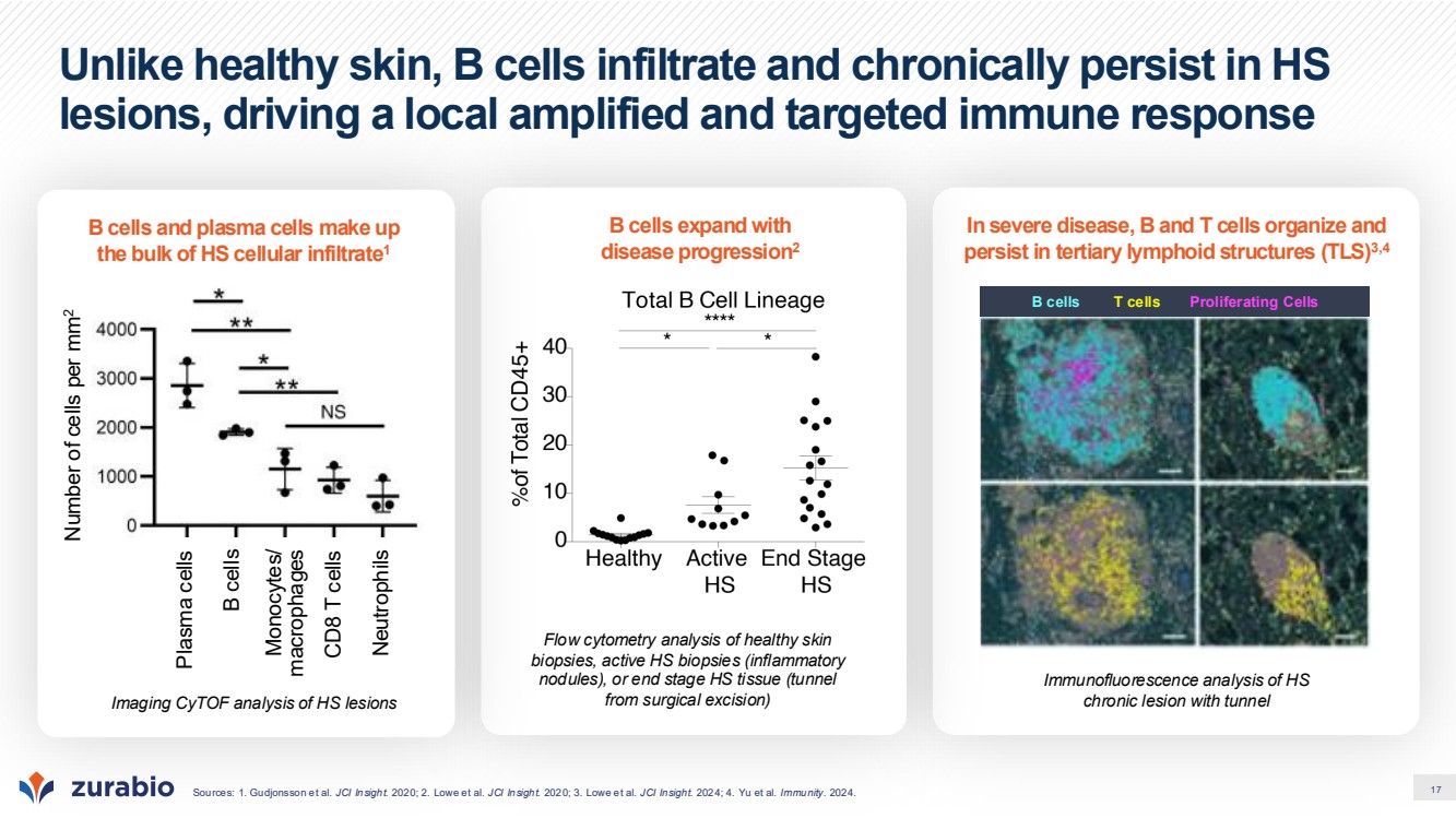
| 18 BAFF is elevated across HS lesion types and predicts treatment resistance In acute and chronic HS lesions, BAFF expression tracks with B cell accumulation, supporting BAFF as a key player in B cell mediated pathology Sources: 1. Yu et al. Immunity. 2024 2. Gudjonsson et al. JCI Insight. 2020; 3. Sabat et al. J Allergy Clin Immunol. 2023; 4. Lowe et al. JCI Insight. 2020; 5. Hambly et al. Br J Dermatol. 2022. Elevated BAFF can retain B cells in the skin Elevated BAFF is a unique feature of HS lesions, which predicts TNFi non-response • BAFF is absent in healthy skin and in psoriasis and dermatitis lesions, and B cells transiently migrate in and out of skin3,4 • BAFF is significantly elevated in all HS lesion types, and correlates with increases in B cells3,4 • Patients who have higher baseline levels of BAFF and B cell counts are less likely to respond to adalimumab4,5 Skin BAFF mRNA (RU) Skin BAFF mRNA (RU) B cells migrate into HS lesions and initiate effector functions1 BAFF promotes B cell accumulation, maturation, and differentiation B and T cells organize into tertiary lymphoid structures that drive chronic, local immune activation1,2 Blood BAFF mRNA (Normalized counts) scars fistulas abscesses nodules P adj=0.005 800 600 400 200 0 |
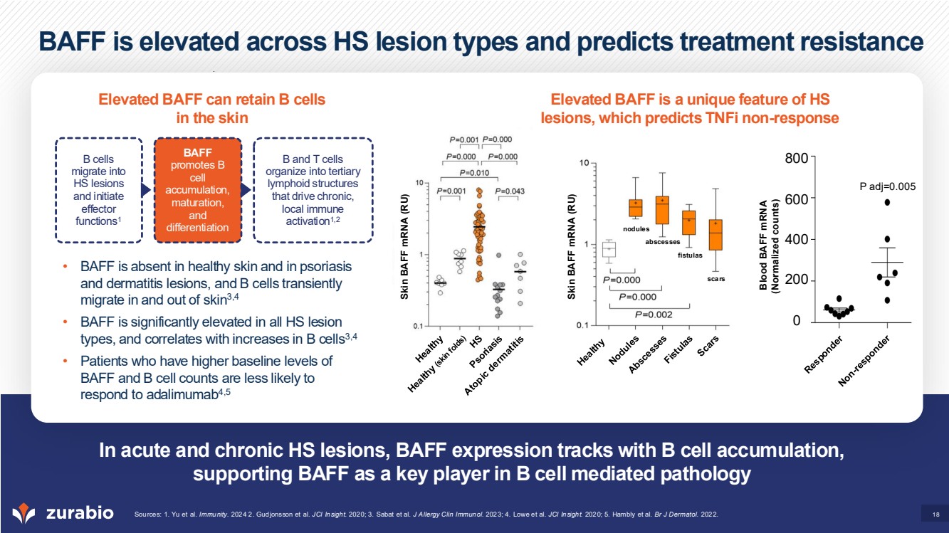
| 19 B cells are believed to drive HS pathology via multiple mechanisms BAFF prolongs B cell survival and promotes several effector functions IL-17 drives a feed-forward, self-sustaining inflammatory cycle Neutrophil recruitment and activation B cell survival Antibody release (IgG, IgA1, IgD) Cytokine and chemokine release T cell activation B cell differentiation into plasma cells IL-17 Th17 Cytokine production (IL-6, TNF, TGFb) BAFF Tertiary Lymphoid B cell Structure |
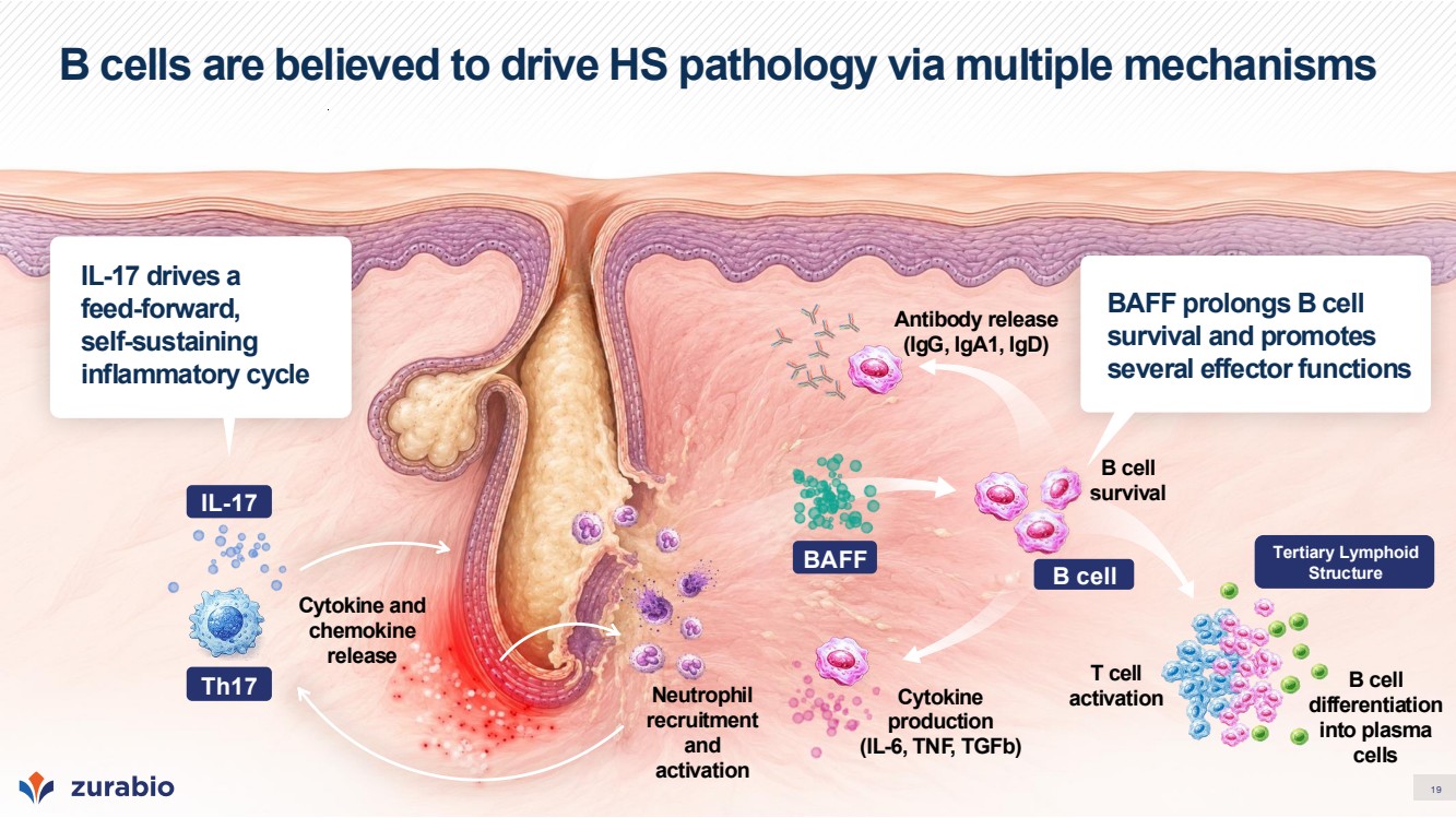
| 20 Three B cell-targeting mechanisms demonstrating HS efficacy signal1,2 Pooled placebo (contemporaneous cohorts) 36% Observed sHiSCR50** Responders at Week 16 by Treatment Arm1,2 Recent clinical data supports the hypothesis that B cells are key drivers of disease rather than bystanders in HS* (*) Data derived from a randomized, placebo-controlled, multi-arm hidradenitis suppurativa platform study (ClinicalTrials.gov Identifier: NCT03827798). Reported results are based on information posted on ClinicalTrials.gov (as of 09-Jan-2026) and are subject to sponsor quality review and potential update. Results are descriptive and not powered for formal cross-arm comparisons. (**) sHiSCR50: ≥50% reduction in abscess and inflammatory nodule count, with no increase in abscesses or draining fistulas, assessed using a simplified lesion-count methodology. Observed response rates; non-responder imputation applied (***) Mean results from 25 mg and 100 mg arm shown Sources: 1. ClinicalTrials.gov. NCT03827798. 2026; 2. Novartis. AAD Annual Meeting. 2024. R W–5 D1 Screening Active Treatment Corresponding Placebo W16 Follow-up period length varies by cohort (4 –84W) PEA EOS Common design applied across 5 cohorts of the Novartis platform study; each cohort tested a different drug against its own randomized placebo arm.1 Platform Study Design 51.6% 72.7% 48.5% 58.6% 46.9% 32.0% ianalumab (VAY736) BAFF-R N=32 remibrutinib (LOU064) BTKi N=33 remibrutinib (LOU064) BTKi N=33 iscalimab (CFZ533) anti-CD-40 N=29 MAS825 IL-1β / IL-18 N=33 LYS006 LPA1 N=27 25 mg 100 mg |
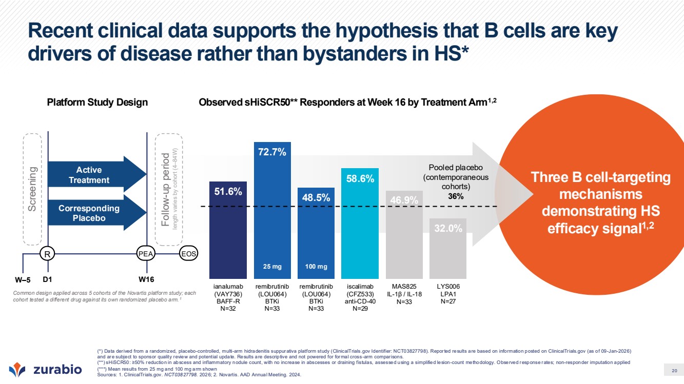
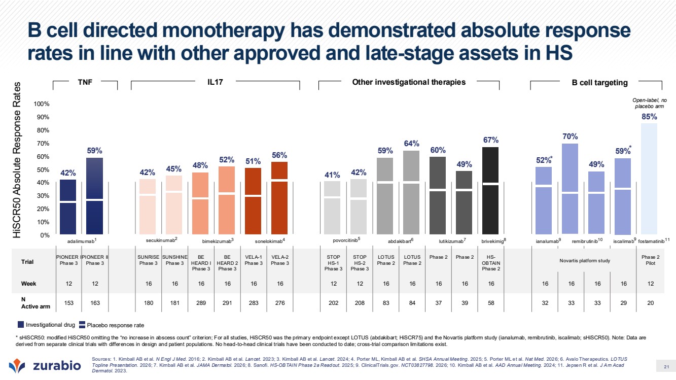
| 21 Trial PIONEER I Phase 3 PIONEER II Phase 3 SUNRISE Phase 3 SUNSHINE Phase 3 BE HEARD I Phase 3 BE HEARD 2 Phase 3 VELA-1 Phase 3 VELA-2 Phase 3 STOP HS-1 Phase 3 STOP HS-2 Phase 3 LOTUS Phase 2 LOTUS Phase 2 Phase 2 Phase 2 HS-OBTAIN Phase 2 Phase 2 Pilot Week 12 12 16 16 16 16 16 16 12 12 16 16 16 16 16 16 16 16 16 12 N Active arm 153 163 180 181 289 291 283 276 202 208 83 84 37 39 58 32 33 33 29 20 B cell directed monotherapy has demonstrated absolute response rates in line with other approved and late-stage assets in HS 42% 59% 42% 45% 48% 52% 51% 56% 41% 42% 59% 64% 60% 49% 67% 52% 70% 49% 59% 85% 0% 10% 20% 30% 40% 50% 60% 70% 80% 90% 100% * * HiSCR50 Absolute Response Rates secukinumab2 adalimumab1 bimekizumab3 sonelokimab4 povorcitinib5 Novartis platform study lutikizumab7 abdakibart6 brivekimig8 ianalumab9 remibrutinib10 fostamatinib11 iscalimab9 Sources: 1. Kimball AB et al. N Engl J Med. 2016; 2. Kimball AB et al. Lancet. 2023; 3. Kimball AB et al. Lancet. 2024; 4. Porter ML, Kimball AB et al. SHSA Annual Meeting. 2025; 5. Porter ML et al. Nat Med. 2026; 6. Avalo Therapeutics. LOTUS Topline Presentation. 2026; 7. Kimball AB et al. JAMA Dermatol. 2026; 8. Sanofi. HS-OBTAIN Phase 2a Readout. 2025; 9. ClinicalTrials.gov. NCT03827798. 2026; 10. Kimball AB et al. AAD Annual Meeting. 2024; 11. Jepsen R et al. J Am Acad Dermatol. 2023. Open-label, no placebo arm TNF IL17 Other investigational therapies B cell targeting * sHiSCR50: modified HiSCR50 omitting the “no increase in abscess count” criterion; For all studies, HiSCR50 was the primary endpoint except LOTUS (abdakibart; HiSCR75) and the Novartis platform study (ianalumab, remibrutinib, iscalimab; sHiSCR50). Note: Data are derived from separate clinical trials with differences in design and patient populations. No head-to-head clinical trials have been conducted to date; cross-trial comparison limitations exist. Investigational drug Placebo response rate |
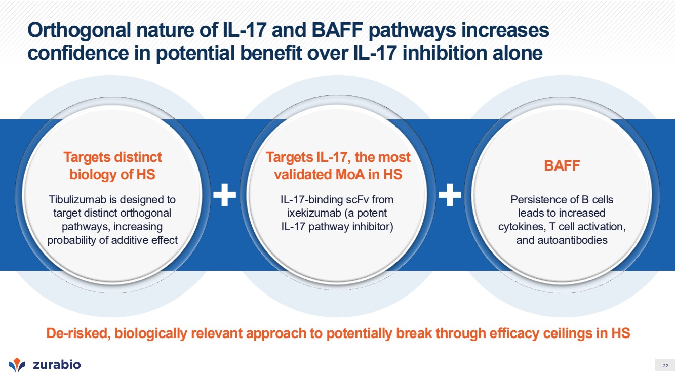
| 22 Orthogonal nature of IL-17 and BAFF pathways increases confidence in potential benefit over IL-17 inhibition alone Targets distinct biology of HS Tibulizumab is designed to target distinct orthogonal pathways, increasing probability of additive effect Targets IL-17, the most validated MoA in HS IL-17-binding scFv from ixekizumab (a potent IL-17 pathway inhibitor) BAFF Persistence of B cells leads to increased cytokines, T cell activation, and autoantibodies De-risked, biologically relevant approach to potentially break through efficacy ceilings in HS |
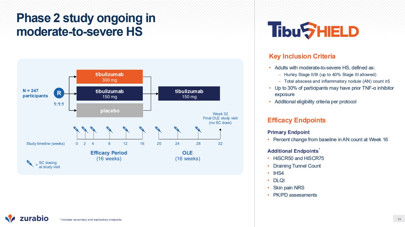
| 23 Phase 2 study ongoing in moderate-to-severe HS Efficacy Endpoints Primary Endpoint • Percent change from baseline in AN count at Week 16 Additional Endpoints* • HiSCR50 and HiSCR75 • Draining Tunnel Count • IHS4 • DLQI • Skin pain NRS • PK/PD assessments Key Inclusion Criteria • Adults with moderate-to-severe HS, defined as: – Hurley Stage II/III (up to 40% Stage III allowed) – Total abscess and inflammatory nodule (AN) count ≥5 • Up to 30% of participants may have prior TNF-α inhibitor exposure • Additional eligibility criteria per protocol Efficacy Period (16 weeks) OLE (16 weeks) Study timeline (weeks) 1:1:1 tibulizumab 150 mg placebo N = 247 participants tibulizumab 300 mg tibulizumab 150 mg 0 2 4 8 12 16 20 24 28 32 Week 32 Final OLE study visit (no SC dose) SC dosing at study visit = * Includes secondary and exploratory endpoints R |
| 24 Systemic Sclerosis (SSc) |
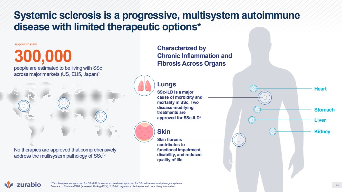
| 25 Systemic sclerosis is a progressive, multisystem autoimmune disease with limited therapeutic options* No therapies are approved that comprehensively address the multisystem pathology of SSc*2 300,000 people are estimated to be living with SSc across major markets (US, EU5, Japan)1 SSc-ILD is a major cause of morbidity and mortality in SSc. Two disease-modifying treatments are approved for SSc-ILD2 Lungs Skin Characterized by Chronic Inflammation and Fibrosis Across Organs Skin fibrosis contributes to functional impairment, disability, and reduced quality of life * Two therapies are approved for SSc-ILD; however, no treatment approved for SSc addresses multiple organ systems. Sources: 1. Clarivate/DRG (accessed 19-Aug-2024); 2. Public regulatory disclosures and prescribing information. Heart Kidney Liver Stomach approximately |
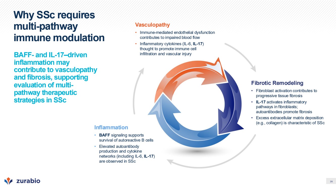
| 26 Why SSc requires multi-pathway immune modulation BAFF- and IL-17–driven inflammation may contribute to vasculopathy and fibrosis, supporting evaluation of multi-pathway therapeutic strategies in SSc Vasculopathy • Immune-mediated endothelial dysfunction contributes to impaired blood flow • Inflammatory cytokines (IL-6, IL-17) thought to promote immune cell infiltration and vascular injury Inflammation • BAFF signaling supports survival of autoreactive B cells • Elevated autoantibody production and cytokine networks (including IL-6, IL-17) are observed in SSc Fibrotic Remodeling • Fibroblast activation contributes to progressive tissue fibrosis • IL-17 activates inflammatory pathways in fibroblasts; autoantibodies promote fibrosis • Excess extracellular matrix deposition (e.g., collagen) is characteristic of SSc |
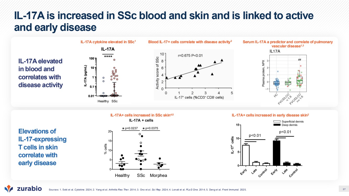
| 27 IL-17A is increased in SSc blood and skin and is linked to active and early disease IL-17A elevated in blood and correlates with disease activity Elevations of IL-17-expressing T cells in skin correlate with early disease IL-17A cytokine elevated in SSc1 Serum IL-17A a predictor and correlate of pulmonary vascular disease1,3 Blood IL-17+ cells correlate with disease activity2 IL-17A+ cells increased in SSc skin4,5 IL-17A+ cells increased in early disease skin2 Sources: 1. Seki et al. Cytokine. 2024; 2. Yang et al. Arthritis Res Ther. 2014; 3. Ono et al. Sci Rep. 2024; 4. Lonati et al. PLoS One. 2014; 5. Deng et al. Front Immunol. 2025. Healthy SSc Morphea Healthy SSc |
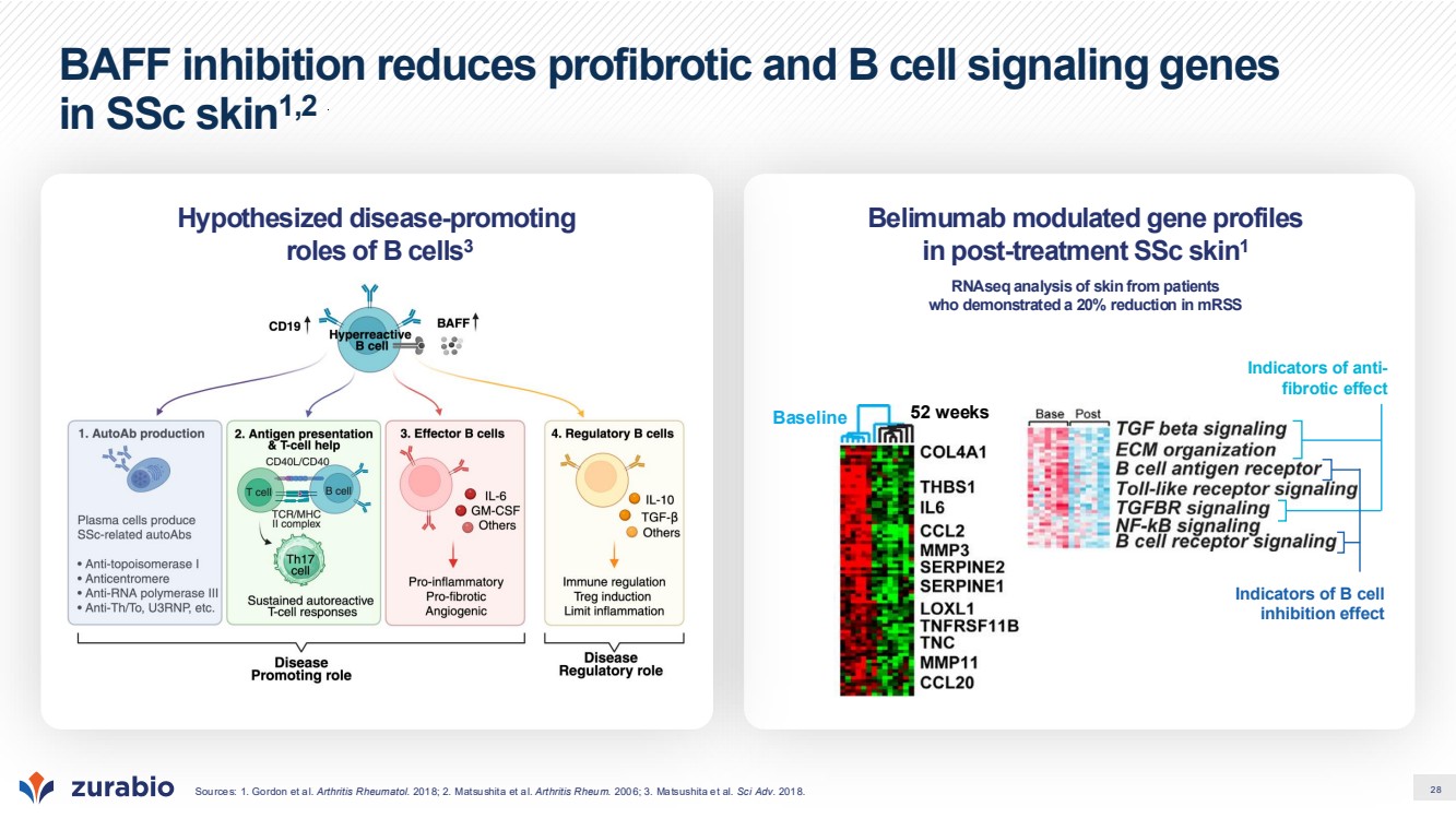
| 28 BAFF inhibition reduces profibrotic and B cell signaling genes in SSc skin1,2 Sources: 1. Gordon et al. Arthritis Rheumatol. 2018; 2. Matsushita et al. Arthritis Rheum. 2006; 3. Matsushita et al. Sci Adv. 2018. Belimumab modulated gene profiles in post-treatment SSc skin1 RNAseq analysis of skin from patients who demonstrated a 20% reduction in mRSS Hypothesized disease-promoting roles of B cells3 Baseline 52 weeks Indicators of anti-fibrotic effect Indicators of B cell inhibition effect |
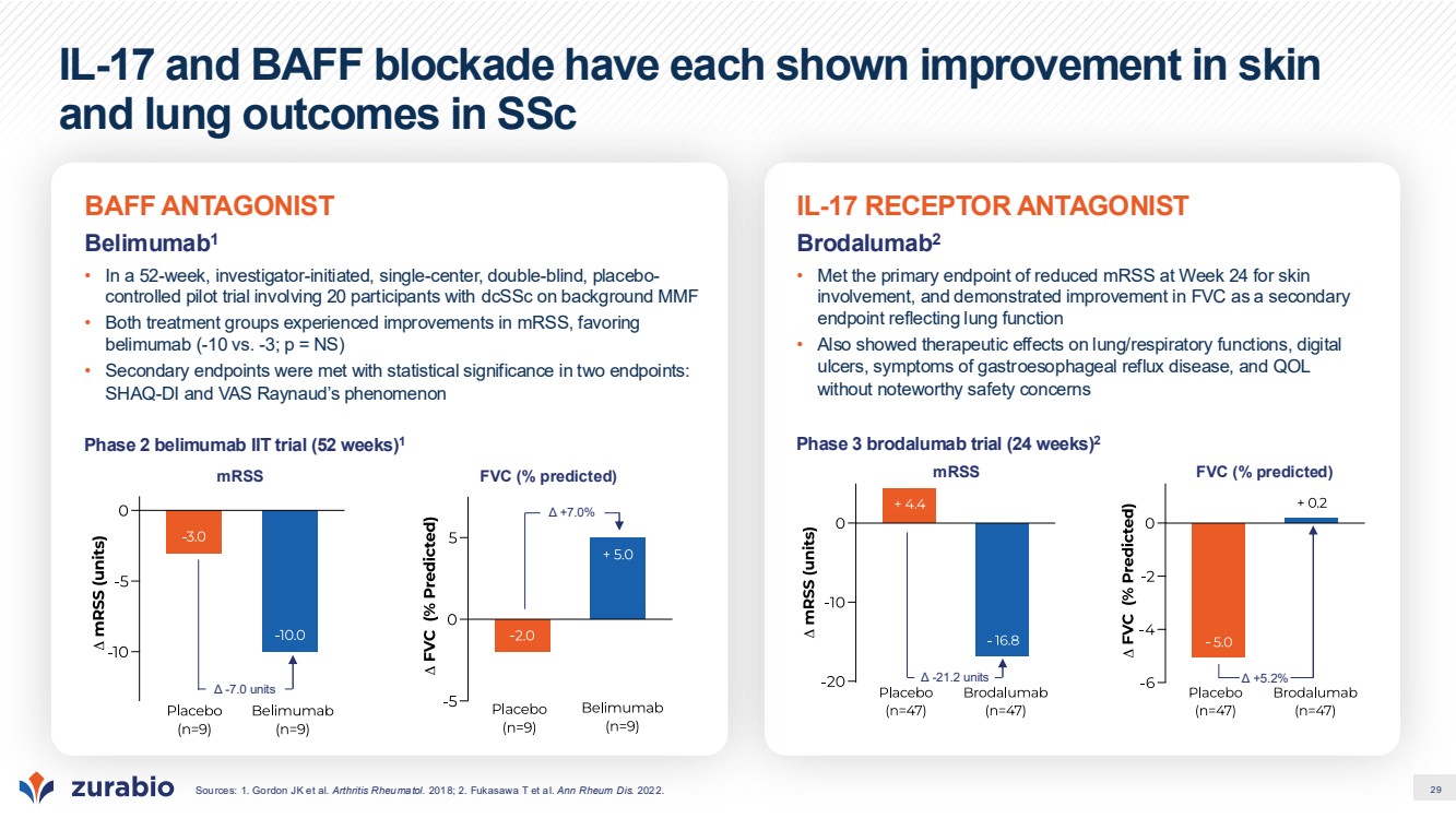
| 29 IL-17 and BAFF blockade have each shown improvement in skin and lung outcomes in SSc Sources: 1. Gordon JK et al. Arthritis Rheumatol. 2018; 2. Fukasawa T et al. Ann Rheum Dis. 2022. IL-17 RECEPTOR ANTAGONIST Brodalumab2 • Met the primary endpoint of reduced mRSS at Week 24 for skin involvement, and demonstrated improvement in FVC as a secondary endpoint reflecting lung function • Also showed therapeutic effects on lung/respiratory functions, digital ulcers, symptoms of gastroesophageal reflux disease, and QOL without noteworthy safety concerns Phase 3 brodalumab trial (24 weeks)2 Δ -21.2 units Δ +5.2% mRSS FVC (% predicted) BAFF ANTAGONIST Belimumab1 • In a 52-week, investigator-initiated, single-center, double-blind, placebo-controlled pilot trial involving 20 participants with dcSSc on background MMF • Both treatment groups experienced improvements in mRSS, favoring belimumab (-10 vs. -3; p = NS) • Secondary endpoints were met with statistical significance in two endpoints: SHAQ-DI and VAS Raynaud’s phenomenon Phase 2 belimumab IIT trial (52 weeks)1 mRSS Δ -7.0 units FVC (% predicted) Δ +7.0% |
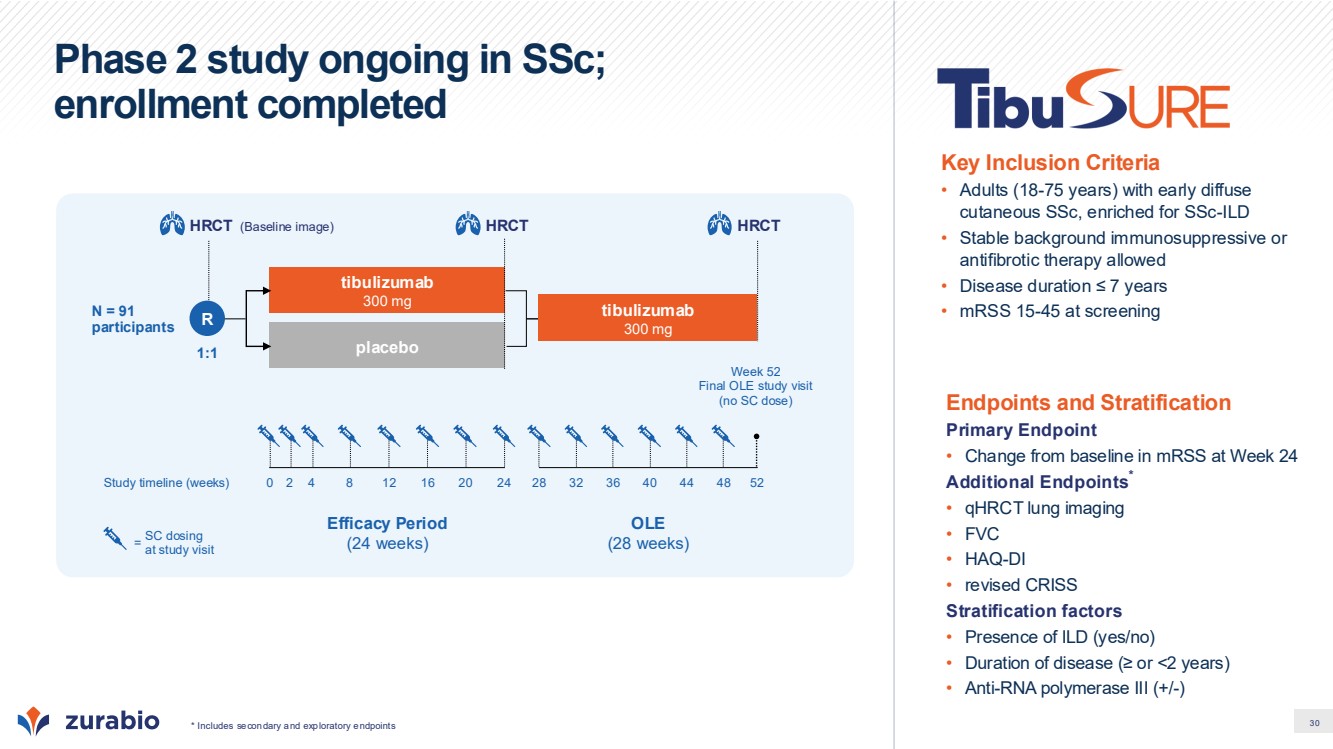
| 30 Phase 2 study ongoing in SSc; enrollment completed * Includes secondary and exploratory endpoints Endpoints and Stratification Primary Endpoint • Change from baseline in mRSS at Week 24 Additional Endpoints* • qHRCT lung imaging • FVC • HAQ-DI • revised CRISS Stratification factors • Presence of ILD (yes/no) • Duration of disease (≥ or <2 years) • Anti-RNA polymerase III (+/-) Key Inclusion Criteria • Adults (18-75 years) with early diffuse cutaneous SSc, enriched for SSc-ILD • Stable background immunosuppressive or antifibrotic therapy allowed • Disease duration ≤ 7 years • mRSS 15-45 at screening Efficacy Period (24 weeks) OLE (28 weeks) 1:1 tibulizumab 300 mg placebo N = 91 participants tibulizumab 300 mg SC dosing at study visit = Study timeline (weeks) 0 52 Week 52 Final OLE study visit (no SC dose) 2 4 8 12 16 20 24 28 32 36 40 44 48 (Baseline image) HRCT R HRCT HRCT |
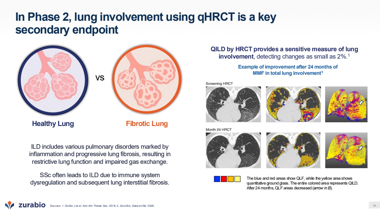
| 31 In Phase 2, lung involvement using qHRCT is a key secondary endpoint Sources: 1. Goldin J et al. Ann Am Thorac Soc. 2018; 2. Zura Bio. Data on file. 2026. ILD includes various pulmonary disorders marked by inflammation and progressive lung fibrosis, resulting in restrictive lung function and impaired gas exchange. SSc often leads to ILD due to immune system dysregulation and subsequent lung interstitial fibrosis. vs Healthy Lung Fibrotic Lung QILD by HRCT provides a sensitive measure of lung involvement, detecting changes as small as 2%.1 Example of improvement after 24 months of MMF in total lung involvement1 The blue and red areas show QLF, while the yellow area shows quantitative ground glass. The entire colored area represents QILD. After 24 months, QLF areas decreased (arrow in B). |
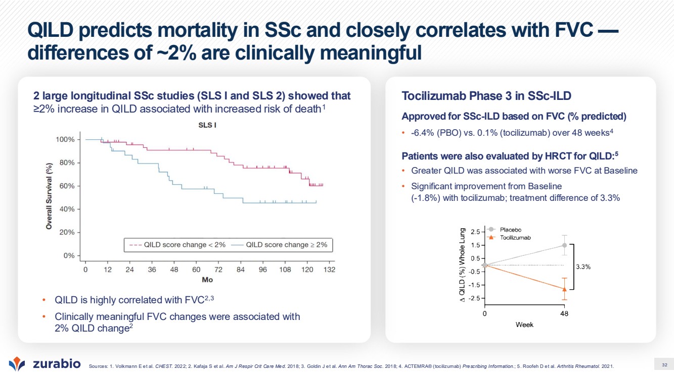
| 32 QILD predicts mortality in SSc and closely correlates with FVC — differences of ~2% are clinically meaningful Sources: 1. Volkmann E et al. CHEST. 2022; 2. Kafaja S et al. Am J Respir Crit Care Med. 2018; 3. Goldin J et al. Ann Am Thorac Soc. 2018; 4. ACTEMRA® (tocilizumab) Prescribing Information.; 5. Roofeh D et al. Arthritis Rheumatol. 2021. 2 large longitudinal SSc studies (SLS I and SLS 2) showed that ≥2% increase in QILD associated with increased risk of death1 Tocilizumab Phase 3 in SSc-ILD Approved for SSc-ILD based on FVC (% predicted) • -6.4% (PBO) vs. 0.1% (tocilizumab) over 48 weeks4 Patients were also evaluated by HRCT for QILD:5 • Greater QILD was associated with worse FVC at Baseline • Significant improvement from Baseline (-1.8%) with tocilizumab; treatment difference of 3.3% • QILD is highly correlated with FVC2,3 • Clinically meaningful FVC changes were associated with 2% QILD change2 |
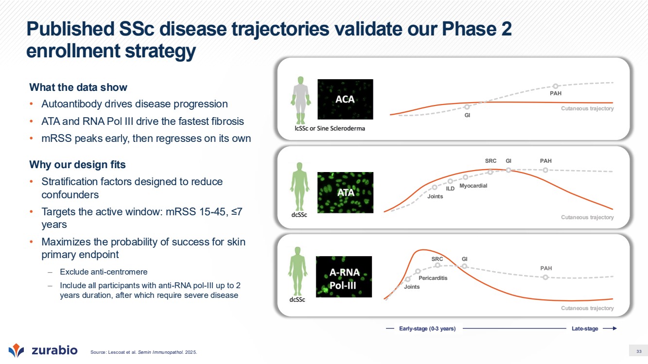
| 33 Published SSc disease trajectories validate our Phase 2 enrollment strategy What the data show • Autoantibody drives disease progression • ATA and RNA Pol III drive the fastest fibrosis • mRSS peaks early, then regresses on its own Why our design fits • Stratification factors designed to reduce confounders • Targets the active window: mRSS 15-45, ≤7 years • Maximizes the probability of success for skin primary endpoint – Exclude anti-centromere – Include all participants with anti-RNA pol-III up to 2 years duration, after which require severe disease Source: Lescoat et al. Semin Immunopathol. 2025. Cutaneous trajectory GI PAH Cutaneous trajectory SRC GI PAH Myocardial ILD Joints Cutaneous trajectory PAH SRC GI Joints Pericarditis Early-stage (0-3 years) Late-stage |
| 34 Polymyalgia Rheumatica (PMR) |
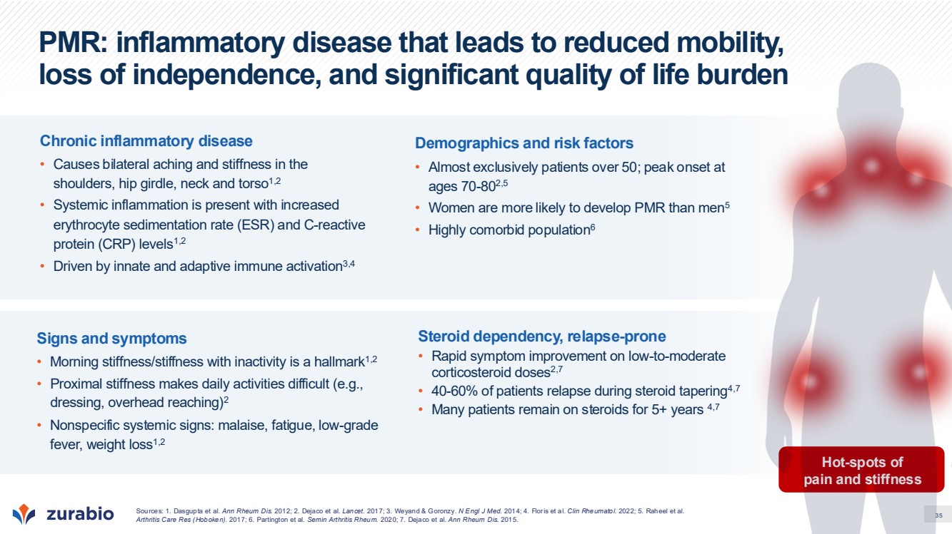
| 35 PMR: inflammatory disease that leads to reduced mobility, loss of independence, and significant quality of life burden Hot-spots of pain and stiffness Chronic inflammatory disease • Causes bilateral aching and stiffness in the shoulders, hip girdle, neck and torso1,2 • Systemic inflammation is present with increased erythrocyte sedimentation rate (ESR) and C-reactive protein (CRP) levels1,2 • Driven by innate and adaptive immune activation3,4 Demographics and risk factors • Almost exclusively patients over 50; peak onset at ages 70-802,5 • Women are more likely to develop PMR than men5 • Highly comorbid population6 Signs and symptoms • Morning stiffness/stiffness with inactivity is a hallmark1,2 • Proximal stiffness makes daily activities difficult (e.g., dressing, overhead reaching)2 • Nonspecific systemic signs: malaise, fatigue, low-grade fever, weight loss1,2 Steroid dependency, relapse-prone • Rapid symptom improvement on low-to-moderate corticosteroid doses2,7 • 40-60% of patients relapse during steroid tapering4,7 • Many patients remain on steroids for 5+ years 4,7 Sources: 1. Dasgupta et al. Ann Rheum Dis. 2012; 2. Dejaco et al. Lancet. 2017; 3. Weyand & Goronzy. N Engl J Med. 2014; 4. Floris et al. Clin Rheumatol. 2022; 5. Raheel et al. Arthritis Care Res (Hoboken). 2017; 6. Partington et al. Semin Arthritis Rheum. 2020; 7. Dejaco et al. Ann Rheum Dis. 2015. |
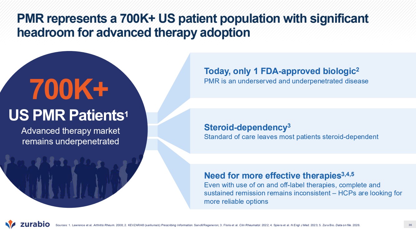
| 36 PMR represents a 700K+ US patient population with significant headroom for advanced therapy adoption Today, only 1 FDA-approved biologic2 PMR is an underserved and underpenetrated disease Need for more effective therapies3,4,5 Even with use of on and off-label therapies, complete and sustained remission remains inconsistent – HCPs are looking for more reliable options Steroid-dependency3 Standard of care leaves most patients steroid-dependent 700K+ US PMR Patients1 Advanced therapy market remains underpenetrated Sources: 1. Lawrence et al. Arthritis Rheum. 2008; 2. KEVZARA® (sarilumab) Prescribing Information. Sanofi/Regeneron; 3. Floris et al. Clin Rheumatol. 2022; 4. Spiera et al. N Engl J Med. 2023; 5. Zura Bio. Data on file. 2026. |
| 37 Current reliance on steroids provides short-term relief but leads to long-term dependence, relapse, and toxicity Steroid-associated toxicities are common (e.g., insomnia, weight gain, hyperglycemia, osteoporosis, and infections) and can be problematic in the PMR age demographic due to high rates of osteoporosis, hypertension, or T2D Median time to permanent glucocorticoid discontinuation Due to high relapse rates during taper, many patients remain on steroids to manage PMR even after 5 years3 Relapse on steroid taper Patients are forced back to higher doses of glucocorticoids with no clear end in sight 50%1 The plan to reduce was difficult. I started reducing by like half a mg a week. The reduction was difficult because the pain kept returning. I couldn’t get away from the pain.” - PMR Patient4 I’m on prednisone, and it does awful things to you. The side effects are really nasty. If there was anything I could take or do to get rid of those side effects, I would do it in a minute. But at this point, apparently, there isn’t anything that my physician knows about.” - PMR Patient4 Sources: 1. Spiera et al. N Engl J Med. 2023; 2. Devauchelle-Pensec et al. Ann Rheum Dis. 2018; 3. Camellino et al. Front Med (Lausanne). 2022; 4. Zura Bio. Data on file. 2026. Over 6 Yrs2 Approx. 37 |
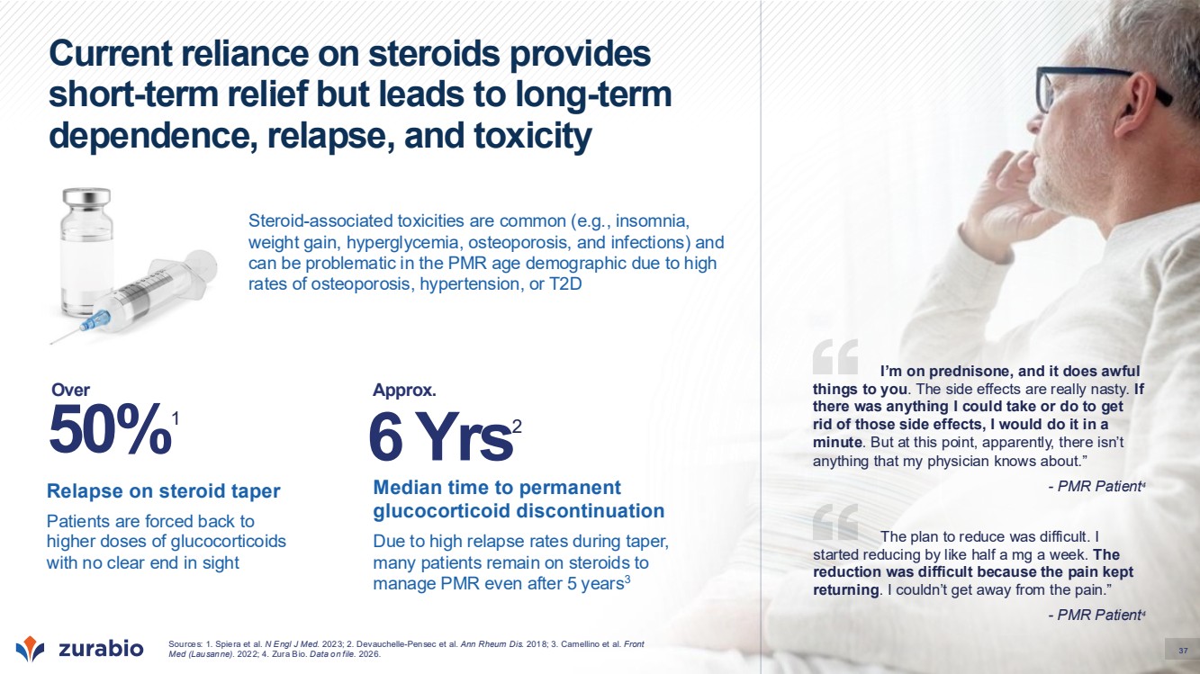
| 38 Targeting BAFF and IL-17 may help address both immune dysregulation and inflammation in PMR Sources: 1. van der Geest et al. Arthritis Rheumatol. 2014; 2. van der Geest et al. Rheumatology (Oxford). 2015; 3. Reitsema et al. Front Immunol. 2022; 4. Ramiro et al. Rheumatol Ther. 2025. B cell homeostasis and trafficking is dysregulated1,2 Circulating B cells: Traffic out of blood during active disease and return in remission1,2 May be acting as antigen presenting cells and cytokine producers (including TNF and IL-6) in sites of inflammation1,2 BAFF correlates with disease activity1,2 Serum BAFF is: Elevated in newly diagnosed PMR1,2 Decreased during remission1,2 Correlated with ESR, CRP, and IL-6 2 ↓may lead to ↑ B cell survival ↑ B cell activation ↑ Cytokine production ↑ Interactions with T cells IL-17 may amplify local and systemic inflammation3,4 IL-17 and IL-17-producing cells (e.g. Th17, MAIT) are: Elevated in circulation3,4 Present in synovial fluid3,4 ↓may lead to ↑ IL-6, IL-8, TNF, and other inflammatory mediators ↑ number and activation of myeloid cells |
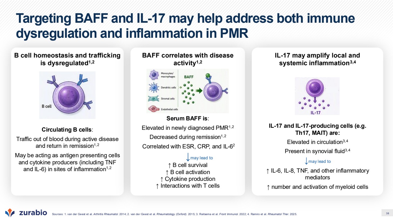
| 39 IL-17 and B cell targeted therapies have shown positive glucocorticoid-sparing effects in PMR * Sustained remission defined as remission that occurred by week 12 and was sustained until week 52, without recurrence of PMR and no new diagnosis of GCA; complete sustained remission defined as sustained remission plus no ESR/CRP elevation at Week 52 ** 52 week results shown from extension phase of BRIDGE-PMR Sources: 1. Stone et al. N Engl J Med. 2026 (REPLENISH); 2. Marsman et al. Lancet Rheumatol. 2021 (BRIDGE-PMR); 3. Bolhuis et al. Lancet Rheumatol. 2023 (BRIDGE-PMR 1-year extension); 4. REDUCE-PMR-1. NCT05533125. Phase 3 REPLENISH trial (52 weeks, N = 381, 1:1:1 ratio) Percentage of patients 41% 28% 41% 25% 20% 5% 0 20 40 60 80 100 Sustained remission Complete sustained remission 89% 43% 52% 86% 43% 54% 78% 24% 34% 0 20 40 60 80 100 Remission by Wk 12 No signs or symptoms, Wks 12–52 No ESR/CRP elevation, Wks 12–52 Secukinumab 300 mg Secukinumab 150 mg Placebo IL-17A inhibitor – relapsing PMR (Phase 3) Secukinumab1 • Met the primary endpoint of sustained remission at Week 52 and all key secondary endpoints • Demonstrated a meaningful glucocorticoid-sparing effect while maintaining disease control • Secukinumab has been filed in the US and EU for approval in PMR Phase 2 BRIDGE-PMR (52 weeks**) and Phase 3 REDUCE-PMR-1 (52 weeks) IITs Glucocorticoid-free remission (PMR-AS <10, No current GC use) 48% 21% 48% 35% 0 20 40 60 80 100 Rituximab Phase 2 Placebo (BRIDGE-PMR) Rituximab Phase 3 Placebo (REDUCE-PMR-1) Anti-CD20 – newly diagnosed PMR (Phase 2 & 3) Rituximab • Anti-CD20 clinical evidence that B cells drive PMR • BRIDGE-PMR (Phase 2 IIT) demonstrated that a single rituximab infusion increased glucocorticoid-free remission at 21 weeks and produced durable benefit through one year2,3 • REDUCE-PMR-1 (Phase 3 IIT) showed trend in favor of rituximab4 Remission Outcomes at Week 52* Components of remission, Weeks 12-52* BRIDGE-PMR (N = 47, 1:1 ratio) REDUCE-PMR-1 (N = 116, 1:1 ratio) |
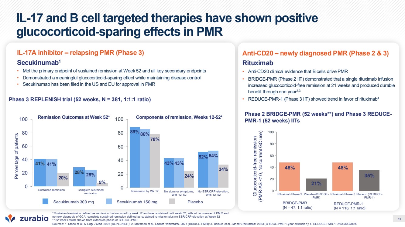
| 40 Despite IL-17 advances, the unmet need for PMR remains significant Source: Stone et al. N Engl J Med. 2026 (REPLENISH). • Glucocorticoids control PMR but cannot be withdrawn: three in four patients on taper alone needed escape or rescue therapy by Week 52 • IL-17 provides real but partial benefit: More than half of participants in secunikumab arms do not reach sustained remission |
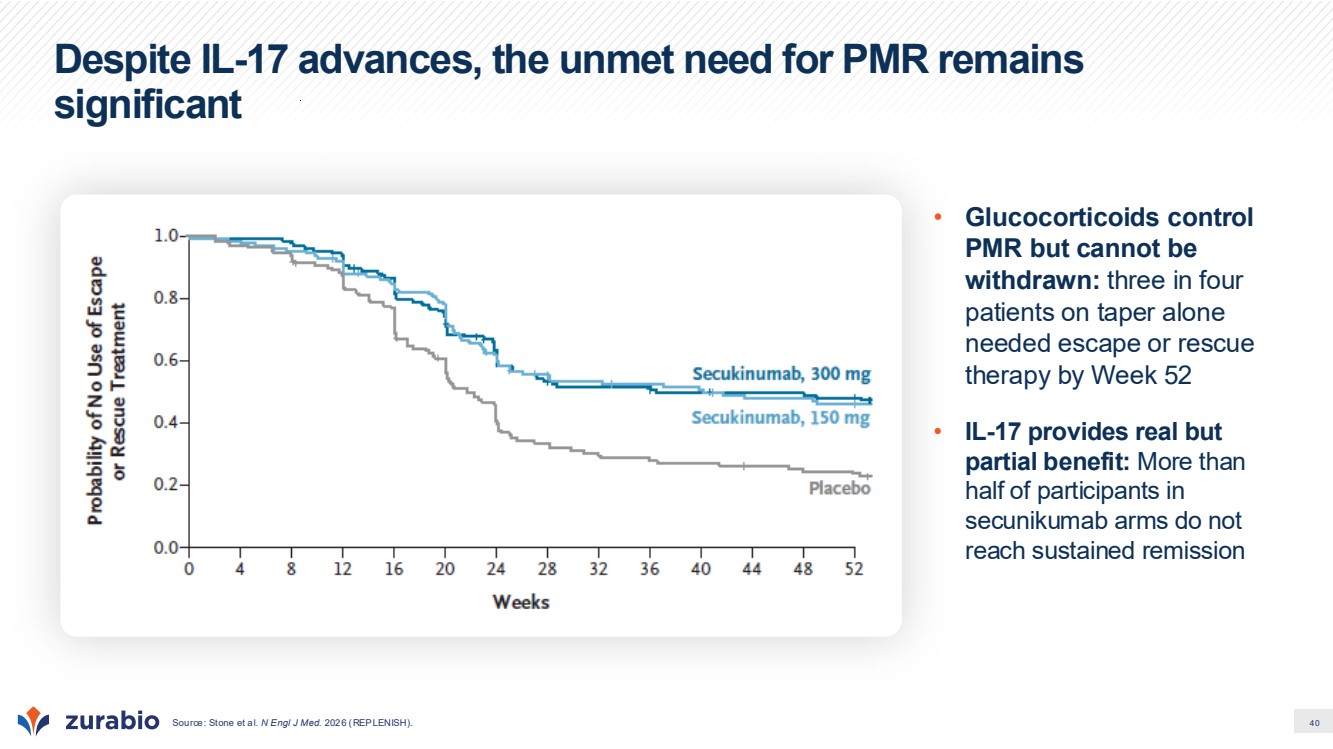
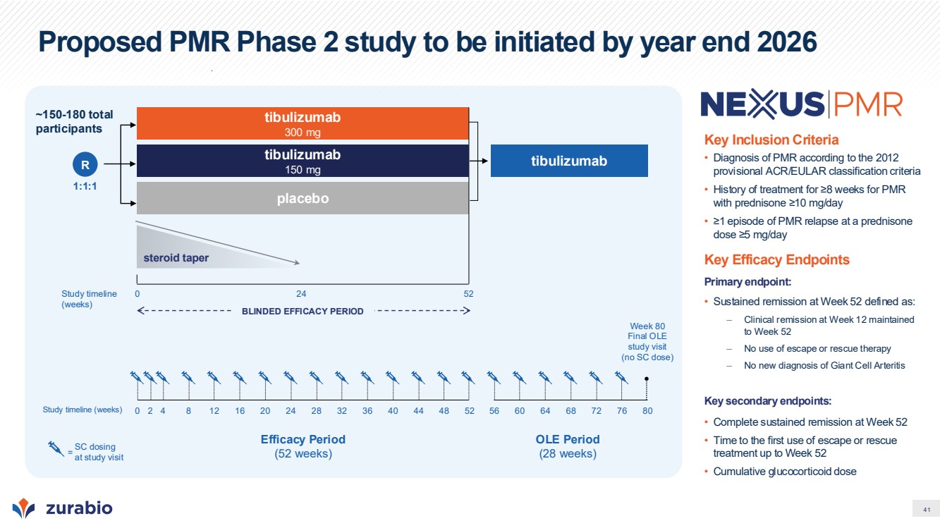
| 41 Proposed PMR Phase 2 study to be initiated by year end 2026 Key Inclusion Criteria • Diagnosis of PMR according to the 2012 provisional ACR/EULAR classification criteria • History of treatment for ≥8 weeks for PMR with prednisone ≥10 mg/day • ≥1 episode of PMR relapse at a prednisone dose ≥5 mg/day Key Efficacy Endpoints Primary endpoint: • Sustained remission at Week 52 defined as: – Clinical remission at Week 12 maintained to Week 52 – No use of escape or rescue therapy – No new diagnosis of Giant Cell Arteritis Key secondary endpoints: • Complete sustained remission at Week 52 • Time to the first use of escape or rescue treatment up to Week 52 • Cumulative glucocorticoid dose tibulizumab 300 mg 1:1:1 R steroid taper placebo Study timeline (weeks) 0 24 52 BLINDED EFFICACY PERIOD tibulizumab 150 mg ~150-180 total participants Efficacy Period (52 weeks) SC dosing at study visit = Study timeline (weeks) 0 52 Week 80 Final OLE study visit (no SC dose) 2 4 8 12 16 20 24 28 32 36 40 44 48 56 60 64 68 72 76 80 OLE Period (28 weeks) tibulizumab |
| 42 Summary & Milestones |

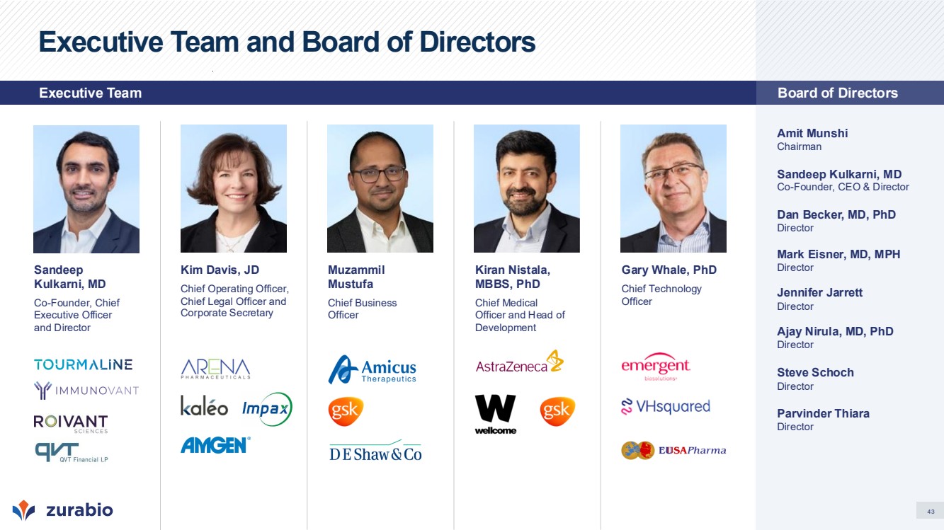
| 43 Executive Team and Board of Directors Amit Munshi Chairman Sandeep Kulkarni, MD Co-Founder, CEO & Director Dan Becker, MD, PhD Director Mark Eisner, MD, MPH Director Jennifer Jarrett Director Ajay Nirula, MD, PhD Director Steve Schoch Director Parvinder Thiara Director Kim Davis, JD Chief Operating Officer, Chief Legal Officer and Corporate Secretary Gary Whale, PhD Chief Technology Officer Kiran Nistala, MBBS, PhD Chief Medical Officer and Head of Development Sandeep Kulkarni, MD Co-Founder, Chief Executive Officer and Director Muzammil Mustufa Chief Business Officer Executive Team Board of Directors |
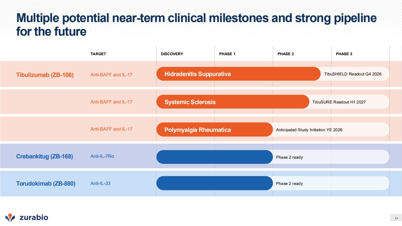
| 44 Multiple potential near-term clinical milestones and strong pipeline for the future TARGET DISCOVERY PHASE 1 PHASE 2 PHASE 3 Tibulizumab (ZB-106) Anti-BAFF and IL-17 Anti-BAFF and IL-17 Anti-BAFF and IL-17 Crebankitug (ZB-168) Anti-IL-7Rα Torudokimab (ZB-880) Anti-IL-33 Hidradenitis Suppurativa Systemic Sclerosis Polymyalgia Rheumatica TibuSURE Readout H1 2027 TibuSHIELD Readout Q4 2026 Anticipated Study Initiation YE 2026 Phase 2 ready Phase 2 ready |
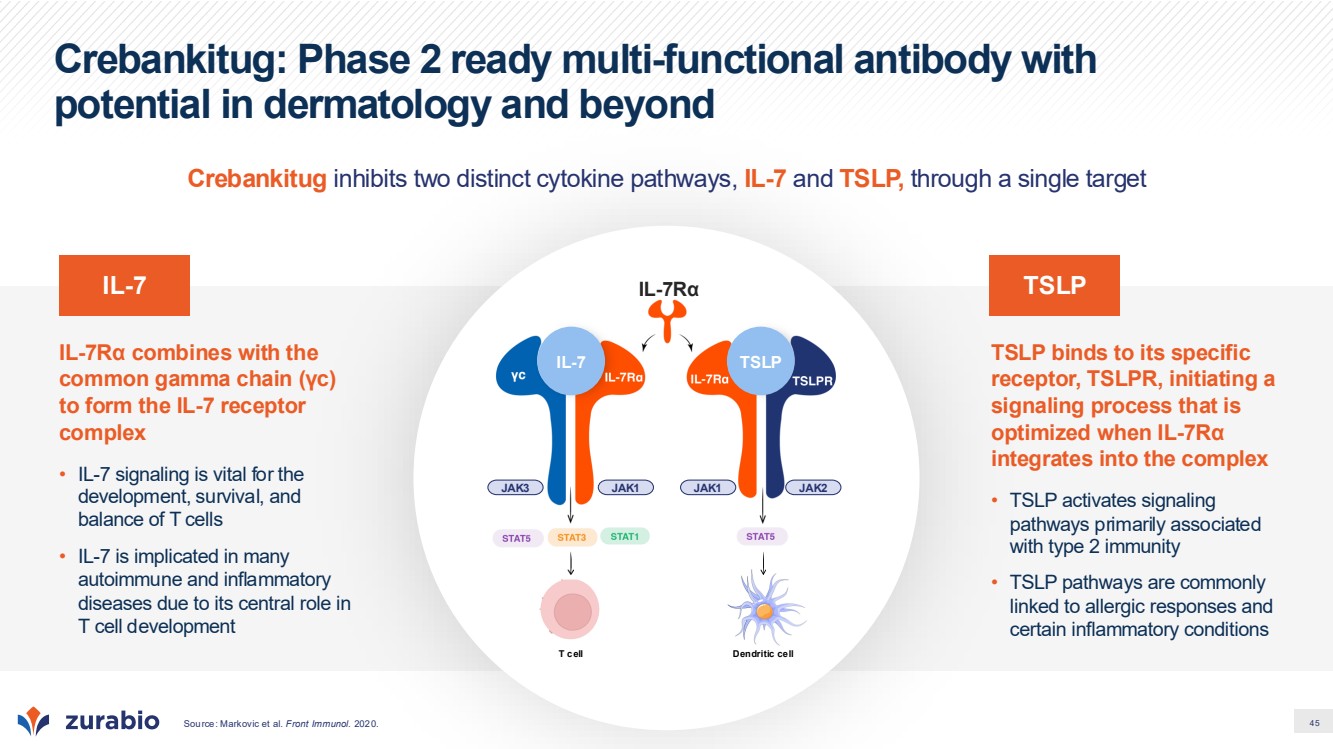
| 45 Crebankitug: Phase 2 ready multi-functional antibody with potential in dermatology and beyond TSLP binds to its specific receptor, TSLPR, initiating a signaling process that is optimized when IL-7Rα integrates into the complex • TSLP activates signaling pathways primarily associated with type 2 immunity • TSLP pathways are commonly linked to allergic responses and certain inflammatory conditions IL-7Rα combines with the common gamma chain (γc) to form the IL-7 receptor complex • IL-7 signaling is vital for the development, survival, and balance of T cells • IL-7 is implicated in many autoimmune and inflammatory diseases due to its central role in T cell development T cell JAK3 JAK1 JAK1 JAK2 IL-7Rα Dendritic cell IL-7 TSLP IL-7 TSLP Crebankitug inhibits two distinct cytokine pathways, IL-7 and TSLP, through a single target Source: Markovic et al. Front Immunol. 2020. |
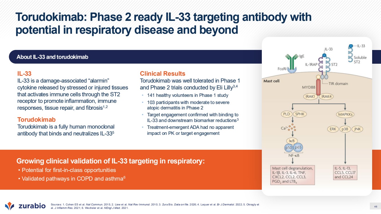
| 46 About IL-33 and torudokimab Growing clinical validation of IL-33 targeting in respiratory: • Potential for first-in-class opportunities • Validated pathways in COPD and asthma6 Torudokimab: Phase 2 ready IL-33 targeting antibody with potential in respiratory disease and beyond Sources: 1. Cohen ES et al. Nat Commun. 2015; 2. Liew et al. Nat Rev Immunol. 2010; 3. Zura Bio. Data on file. 2026; 4. Laquer et al. Br J Dermatol. 2022; 5. Okragly et al. J Inflamm Res. 2021; 6. Wechsler et al. NEngl J Med. 2021. IL-33 IL-33 is a damage-associated “alarmin” cytokine released by stressed or injured tissues that activates immune cells through the ST2 receptor to promote inflammation, immune responses, tissue repair, and fibrosis1,2 Torudokimab Torudokimab is a fully human monoclonal antibody that binds and neutralizes IL-335 Clinical Results Torudokimab was well tolerated in Phase 1 and Phase 2 trials conducted by Eli Lilly3,4 • 141 healthy volunteers in Phase 1 study • 103 participants with moderate to severe atopic dermatitis in Phase 2 • Target engagement confirmed with binding to IL-33 and downstream biomarker reductions3 • Treatment-emergent ADA had no apparent impact on PK or target engagement |
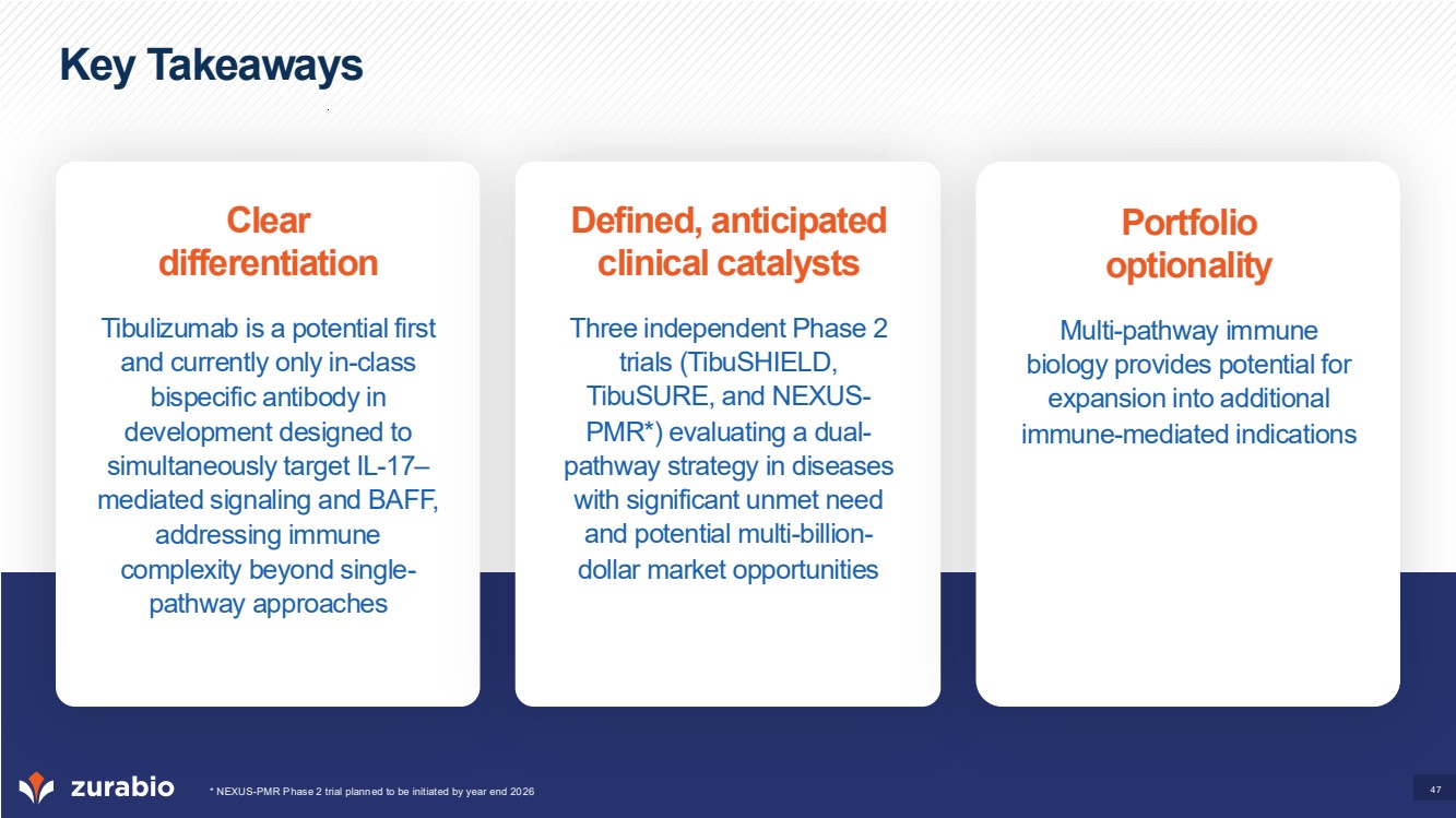
| 47 Key Takeaways Clear differentiation Tibulizumab is a potential first and currently only in-class bispecific antibody in development designed to simultaneously target IL-17– mediated signaling and BAFF, addressing immune complexity beyond single-pathway approaches Defined, anticipated clinical catalysts Three independent Phase 2 trials (TibuSHIELD, TibuSURE, and NEXUS-PMR*) evaluating a dual-pathway strategy in diseases with significant unmet need and potential multi-billion-dollar market opportunities Portfolio optionality Multi-pathway immune biology provides potential for expansion into additional immune-mediated indications * NEXUS-PMR Phase 2 trial planned to be initiated by year end 2026 |
| 48 Appendix |

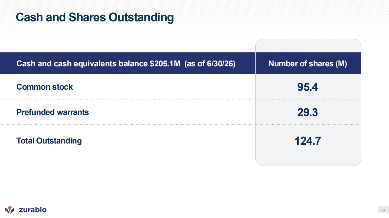
| 49 Cash and Shares Outstanding Cash and cash equivalents balance $205.1M (as of 6/30/26) Number of shares (M) Common stock 95.4 Prefunded warrants 29.3 Total Outstanding 124.7 |
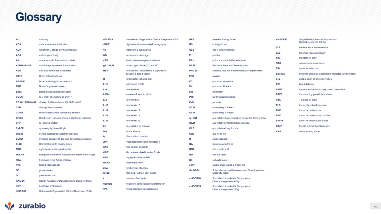
| 50 Glossary Ab antibody ACA anti-centromere antibodies ACR American College of Rheumatology ADA anti-drug antibody AN abscess and inflammatory nodule A-RNA-Pol-III anti-RNA polymerase 3 antibodies ATA anti-topoisomerase antibodies BAFF B cell activating factor BAFF-R B cell activating factor receptor BTK Bruton’s tyrosine kinase BTKi Bruton’s tyrosine kinase inhibitors CCL17 C-C motif chemokine ligand 17 CD3/8/19/20/40/45 cluster of differentiation 3/8/19/20/40/45 CFB change from baseline COPD chronic obstructive pulmonary disease CRISS Combined Response Index in Systemic Sclerosis CRP C-reactive protein CyTOF cytometry by time-of-flight dcSSc diffuse cutaneous systemic sclerosis DLCO diffusing capacity of the lung for carbon monoxide DLQI Dermatology Life Quality Index ESR erythrocyte sedimentation rate EULAR European Alliance of Associations for Rheumatology FDA Food and Drug Administration FVC forced vital capacity GC glucocorticoid GI gastrointestinal HAQ-DI Health Assessment Questionnaire Disability Index HCP healthcare professional HiSCR50 Hidradenitis Suppurativa Clinical Response ≥50% HiSCR75 Hidradenitis Suppurativa Clinical Response ≥75% HRCT high-resolution computed tomography HS hidradenitis suppurativa I&D incision and drainage lcSSc limited cutaneous systemic sclerosis IgA1, D, G immunoglobulin A1, D, and G IHS4 International Hidradenitis Suppurativa Severity Score System IIT investigator-initiated trial IL-1β interleukin 1 beta IL-6 interleukin 6 IL-7Rα interleukin 7 receptor alpha IL-8 interleukin 8 IL-10 interleukin 10 IL-17 interleukin 17 IL-18 interleukin 18 IL-33 interleukin 33 ILD interstitial lung disease JAK Janus kinase KD dissociation constant LPA1 lysophosphatidic acid receptor 1 mAb monoclonal antibody MAIT Mucosal-associated invariant T cells MMF mycophenolate mofetil mRNA messenger RNA MoA mechanism of action mRSS Modified Rodnan Skin Score N number of subjects NETosis neutrophil extracellular trap formation NPX normalized protein expression NRS Numeric Rating Scale NS not significant OLE open-label extension P p-value PAH pulmonary arterial hypertension PASI Psoriasis Area and Severity Index PASI 90 Psoriasis Area and Severity Index 90% improvement PBO placebo PD pharmacodynamics PK pharmacokinetics pM picomolar PMR polymyalgia rheumatica PsO psoriasis Q2W once every 2 weeks Q4W once every 4 weeks qHRCT quantitative high-resolution computed tomography QILD quantitative interstitial lung disease QLF quantitative lung fibrosis QOL quality of life R randomization RA rheumatoid arthritis RNA ribonucleic acid RU relative units SC subcutaneous scFv single-chain variable fragment SHAQ-DI Scleroderma Health Assessment Questionnaire– Disability Index sHiSCR50 Simplified Hidradenitis Suppurativa Clinical Response (50%) sHiSCR75 Simplified Hidradenitis Suppurativa Clinical Response (75%) sHiSCR90 Simplified Hidradenitis Suppurativa Clinical Response (90%) SLE systemic lupus erythematosus SLS Scleroderma Lung Study SoC standard of care SRC scleroderma renal crisis SSc systemic sclerosis SSc-ILD systemic sclerosis-associated interstitial lung disease ST2 suppression of tumorigenicity 2 T2D type 2 diabetes TARC thymus and activation-regulated chemokine TGFβ transforming growth factor beta Th17 T helper 17 cells TLS tertiary lymphoid structure TNF tumor necrosis factor TNFi tumor necrosis factor inhibitor TNF-α tumor necrosis factor alpha TSLP thymic stromal lymphopoietin VAS visual analog scale |
| Nasdaq: ZURA | www.zurabio.com |